 Movie
Movie Controller


+ Open data

- Basic information
| Entry | Database: PDB / ID: 9j60 | ||||||||||||||||||||||||
|---|---|---|---|---|---|---|---|---|---|---|---|---|---|---|---|---|---|---|---|---|---|---|---|---|---|
| Title | Cryo-EM structure of the rice isoamylase ISA1 dimer | ||||||||||||||||||||||||
 Components Components | Isoamylase 1, chloroplastic | ||||||||||||||||||||||||
 Keywords Keywords | HYDROLASE / isoamylase ISA1-ISA2 | ||||||||||||||||||||||||
| Function / homology |  Function and homology information Function and homology informationisoamylase complex / chloroplast isoamylase complex / isoamylase / isoamylase activity / amylopectin biosynthetic process / starch biosynthetic process / starch catabolic process Similarity search - Function | ||||||||||||||||||||||||
| Biological species |  | ||||||||||||||||||||||||
| Method | ELECTRON MICROSCOPY / single particle reconstruction / cryo EM / Resolution: 2.7 Å | ||||||||||||||||||||||||
 Authors Authors | Guan, Z.Y. / Yan, J.J. | ||||||||||||||||||||||||
| Funding support |  China, 1items China, 1items
| ||||||||||||||||||||||||
 Citation Citation | Journal: Nat Commun / Year: 2025 Title: Amylopectin branch trimming and biosynthesis elucidated by the rice isoamylase ISA1-ISA2 heterocomplex. Authors: Rong Fan / Zeyuan Guan / Guanghong Zhou / Xi Yang / Fei Zhang / Menglong Wu / Xuecui Wang / Jian Liu / Pei Chen / Yanjun Liu / Delin Zhang / Ping Yin / Junjie Yan / Abstract: Amylopectin, the primary form of starch in plant leaves, seeds and tubers, features a tree-like architecture with branched glucose chains. Excess branches result in the formation of soluble ...Amylopectin, the primary form of starch in plant leaves, seeds and tubers, features a tree-like architecture with branched glucose chains. Excess branches result in the formation of soluble phytoglycogen instead of starch granules. In higher plants and green algae, the debranching enzyme isoamylase ISA1 forms either homomultimer or hetero-multimer with ISA2 to facilitate branch trimming and starch granule formation, but the molecular basis remains largely unknown. In this study, we reconstitute the rice OsISA1-ISA2 complex in vitro and determine the cryo-EM structures of the OsISA1 homodimer, as well as the malto-oligosaccharide (MOS)-free and MOS-bound OsISA1-ISA2 heterocomplex. The OsISA1 dimer shows a tail-to-tail rod-like architecture, whereas the OsISA1-ISA2 complex mainly exhibits as a trimer, with OsISA2 flanking on the N-terminal segments of the dimeric OsISA1. Combined with comprehensive biochemical analyses, these structural data elucidate the organization of the ISA1-ISA2 heterocomplex in higher plants and demonstrate how ISA1 and ISA2 cooperate during amylopectin biosynthesis. | ||||||||||||||||||||||||
| History |
|
- Structure visualization
| Structure viewer | Molecule: MolmilJmol/JSmol |
|---|
- Downloads & links
-Download
| PDBx/mmCIF format | 9j60.cif.gz | 497.3 KB | Display | PDBx/mmCIF format |
|---|---|---|---|---|
| PDB format | pdb9j60.ent.gz | 415 KB | Display | PDB format |
| PDBx/mmJSON format | 9j60.json.gz | Tree view | PDBx/mmJSON format | |
| Others |  Other downloads Other downloads |
-Validation report
| Arichive directory | https://data.pdbj.org/pub/pdb/validation_reports/j6/9j60ftp://data.pdbj.org/pub/pdb/validation_reports/j6/9j60 | HTTPS FTP |
|---|
-Related structure data
| Related structure data |  61158MC  9j6xC 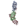 9lfnC M: map data used to model this data C: citing same article ( |
|---|---|
| Similar structure data |


-Links
 PDBj
PDBj

- Assembly
| Deposited unit | 
|
|---|---|
| 1 |
|
-Components
| #1: Protein | Mass: 87281.594 Da / Num. of mol.: 2 Source method: isolated from a genetically manipulated source Source: (gene. exp.) Gene: ISA1, ISA, SU1, Os08g0520900, LOC_Os08g40930 / Production host:  Has protein modification | N | |
|---|
-Experimental details
-Experiment
| Experiment | Method: ELECTRON MICROSCOPY |
|---|---|
| EM experiment | Aggregation state: PARTICLE / 3D reconstruction method: single particle reconstruction |
- Sample preparation
| Component | Name: ISA1 dimer / Type: COMPLEX / Entity ID: all / Source: RECOMBINANT |
|---|---|
| Source (natural) | Organism: |
| Source (recombinant) | Organism: |
| Buffer solution | pH: 8 |
| Specimen | Conc.: 0.75 mg/ml / Embedding applied: NO / Shadowing applied: NO / Staining applied: NO / Vitrification applied: YES |
| Vitrification | Cryogen name: ETHANE |
- Electron microscopy imaging
| Experimental equipment |  Model: Titan Krios / Image courtesy: FEI Company |
|---|---|
| Microscopy | Model: TFS KRIOS |
| Electron gun | Electron source:  FIELD EMISSION GUN / Accelerating voltage: 300 kV / Illumination mode: FLOOD BEAM FIELD EMISSION GUN / Accelerating voltage: 300 kV / Illumination mode: FLOOD BEAM |
| Electron lens | Mode: BRIGHT FIELD / Nominal defocus max: 1600 nm / Nominal defocus min: 1200 nm |
| Image recording | Electron dose: 50 e/Å2 / Film or detector model: GATAN K3 BIOQUANTUM (6k x 4k) |
- Processing
| EM software | Name: PHENIX / Category: model refinement | ||||||||||||||||||||||||
|---|---|---|---|---|---|---|---|---|---|---|---|---|---|---|---|---|---|---|---|---|---|---|---|---|---|
| CTF correction | Type: NONE | ||||||||||||||||||||||||
| Symmetry | Point symmetry: C2 (2 fold cyclic) | ||||||||||||||||||||||||
| 3D reconstruction | Resolution: 2.7 Å / Resolution method: FSC 0.143 CUT-OFF / Num. of particles: 104741 / Symmetry type: POINT | ||||||||||||||||||||||||
| Refinement | Highest resolution: 2.7 Å Stereochemistry target values: REAL-SPACE (WEIGHTED MAP SUM AT ATOM CENTERS) | ||||||||||||||||||||||||
| Refine LS restraints |
|
