+ Open data
- Basic information
| Entry | Database: PDB / ID: 9j3c | ||||||
|---|---|---|---|---|---|---|---|
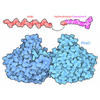
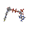
| Title | Cryo-EM structure of NAT10 with Co-enzyme A | ||||||
 Components Components | RNA cytidine acetyltransferase | ||||||
 Keywords Keywords | TRANSFERASE / NAT10 / acyltransferase / N4-acetylcytidine | ||||||
| Function / homology |  Function and homology information Function and homology informationmRNA N-acetyltransferase activity / tRNA wobble cytosine modification / tRNA cytidine N4-acetyltransferase activity / rRNA acetylation involved in maturation of SSU-rRNA / 18S rRNA cytidine N-acetyltransferase activity / tRNA acetylation / rRNA modification / regulation of centrosome duplication / N-acetyltransferase activity / telomerase holoenzyme complex ...mRNA N-acetyltransferase activity / tRNA wobble cytosine modification / tRNA cytidine N4-acetyltransferase activity / rRNA acetylation involved in maturation of SSU-rRNA / 18S rRNA cytidine N-acetyltransferase activity / tRNA acetylation / rRNA modification / regulation of centrosome duplication / N-acetyltransferase activity / telomerase holoenzyme complex / protein acetylation / rRNA modification in the nucleus and cytosol / negative regulation of telomere maintenance via telomerase / DNA polymerase binding / Transferases; Acyltransferases; Transferring groups other than aminoacyl groups / positive regulation of translation / small-subunit processome / regulation of translation / ribosomal small subunit biogenesis / midbody / tRNA binding / chromosome, telomeric region / nucleolus / RNA binding / nucleoplasm / ATP binding / nucleus / membrane Similarity search - Function | ||||||
| Biological species |  Homo sapiens (human) Homo sapiens (human) | ||||||
| Method | ELECTRON MICROSCOPY / single particle reconstruction / cryo EM / Resolution: 2.9 Å | ||||||
 Authors Authors | Jiang, Y. / Xia, J. | ||||||
| Funding support | 1items
| ||||||
 Citation Citation |  Journal: Drug Resist Updat / Year: 2025 Journal: Drug Resist Updat / Year: 2025Title: Targeting NAT10 attenuates homologous recombination via destabilizing DNA:RNA hybrids and overcomes PARP inhibitor resistance in cancers. Authors: Zhu Xu / Mingming Zhu / Longpo Geng / Jun Zhang / Jing Xia / Qiang Wang / Hongda An / Anliang Xia / Yuanyuan Yu / Shihan Liu / Junjie Tong / Wei-Guo Zhu / Yiyang Jiang / Beicheng Sun /  Abstract: AIMS: RNA metabolism has been extensively studied in DNA double-strand break (DSB) repair. The RNA acetyltransferase N-acetyltransferase 10 (NAT10)-mediated N4-acetylcytidine (ac4C) modification in ...AIMS: RNA metabolism has been extensively studied in DNA double-strand break (DSB) repair. The RNA acetyltransferase N-acetyltransferase 10 (NAT10)-mediated N4-acetylcytidine (ac4C) modification in DSB repair remains largely elusive. In this study, we aim to decipher the role for ac4C modification by NAT10 in DSB repair in hepatocellular carcinoma (HCC). METHODS: Laser micro-irradiation and chromatin immunoprecipitation (ChIP) were used to assess the accumulation of ac4C modification and NAT10 at DSB sites. Cryo-electron microscopy (cryo-EM) was used ...METHODS: Laser micro-irradiation and chromatin immunoprecipitation (ChIP) were used to assess the accumulation of ac4C modification and NAT10 at DSB sites. Cryo-electron microscopy (cryo-EM) was used to determine the structures of NAT10 in complex with its inhibitor, remodelin. Hepatocyte-specific deletion of NAT10 mouse models were adopted to detect the effects of NAT10 on HCC progression. Subcutaneous xenograft, human HCC organoid and patient-derived xenograft (PDX) model were exploited to determine the therapy efficiency of the combination of a poly (ADP-ribose) polymerase 1 (PARP1) inhibitor (PARPi) and remodelin. RESULTS: NAT10 promptly accumulates at DSB sites, where it executes ac4C modification on RNAs at DNA:RNA hybrids dependent on PARP1. This in turn enhances the stability of DNA:RNA hybrids and ...RESULTS: NAT10 promptly accumulates at DSB sites, where it executes ac4C modification on RNAs at DNA:RNA hybrids dependent on PARP1. This in turn enhances the stability of DNA:RNA hybrids and promotes homologous recombination (HR) repair. The ablation of NAT10 curtails HCC progression. Furthermore, the cryo-EM yields a remarkable 2.9 angstroms resolution structure of NAT10-remodelin, showcasing a C2 symmetric architecture. Remodelin treatment significantly enhanced the sensitivity of HCC cells to a PARPi and targeting NAT10 also restored sensitivity to a PARPi in ovarian and breast cancer cells that had developed resistance. CONCLUSION: Our study elucidated the mechanism of NAT10-mediated ac4C modification in DSB repair, revealing that targeting NAT10 confers synthetic lethality to PARP inhibition in HCC. Our findings ...CONCLUSION: Our study elucidated the mechanism of NAT10-mediated ac4C modification in DSB repair, revealing that targeting NAT10 confers synthetic lethality to PARP inhibition in HCC. Our findings suggest that co-inhibition of NAT10 and PARP1 is an effective novel therapeutic strategy for patients with HCC and have the potential to overcome PARPi resistance. | ||||||
| History |
|
- Structure visualization
| Structure viewer | Molecule:  Molmil Molmil Jmol/JSmol Jmol/JSmol |
|---|
- Downloads & links
- Download
| PDBx/mmCIF format |  9j3c.cif.gz 9j3c.cif.gz | 390.4 KB | Display |  PDBx/mmCIF format PDBx/mmCIF format |
|---|---|---|---|---|
| PDB format |  pdb9j3c.ent.gz pdb9j3c.ent.gz | 249.3 KB | Display |  PDB format PDB format |
| PDBx/mmJSON format |  9j3c.json.gz 9j3c.json.gz | Tree view |  PDBx/mmJSON format PDBx/mmJSON format | |
| Others |  Other downloads Other downloads |
-Validation report
| Summary document |  9j3c_validation.pdf.gz 9j3c_validation.pdf.gz | 1.3 MB | Display |  wwPDB validaton report wwPDB validaton report |
|---|---|---|---|---|
| Full document |  9j3c_full_validation.pdf.gz 9j3c_full_validation.pdf.gz | 1.4 MB | Display | |
| Data in XML |  9j3c_validation.xml.gz 9j3c_validation.xml.gz | 54.8 KB | Display | |
| Data in CIF |  9j3c_validation.cif.gz 9j3c_validation.cif.gz | 78.6 KB | Display | |
| Arichive directory |  https://data.pdbj.org/pub/pdb/validation_reports/j3/9j3c https://data.pdbj.org/pub/pdb/validation_reports/j3/9j3c ftp://data.pdbj.org/pub/pdb/validation_reports/j3/9j3c ftp://data.pdbj.org/pub/pdb/validation_reports/j3/9j3c | HTTPS FTP |
-Related structure data
| Related structure data |  61113MC M: map data used to model this data C: citing same article ( |
|---|---|
| Similar structure data | Similarity search - Function & homology  F&H Search F&H Search |
- Links
Links
- Assembly
| Deposited unit | 
|
|---|---|
| 1 |
|
- Components
| #1: Protein | Mass: 103855.844 Da / Num. of mol.: 2 Source method: isolated from a genetically manipulated source Source: (gene. exp.)  Homo sapiens (human) / Gene: NAT10, ALP, KIAA1709 / Production host: Homo sapiens (human) / Gene: NAT10, ALP, KIAA1709 / Production host:  Homo sapiens (human) Homo sapiens (human)References: UniProt: Q9H0A0, Transferases; Acyltransferases; Transferring groups other than aminoacyl groups #2: Chemical | Has ligand of interest | Y | Has protein modification | N | |
|---|
-Experimental details
-Experiment
| Experiment | Method: ELECTRON MICROSCOPY |
|---|---|
| EM experiment | Aggregation state: PARTICLE / 3D reconstruction method: single particle reconstruction |
- Sample preparation
| Component | Name: NAT10 with Co-enzyme A / Type: COMPLEX / Entity ID: #1 / Source: RECOMBINANT |
|---|---|
| Molecular weight | Experimental value: NO |
| Source (natural) | Organism:  Homo sapiens (human) Homo sapiens (human) |
| Source (recombinant) | Organism:  Homo sapiens (human) Homo sapiens (human) |
| Buffer solution | pH: 7.4 / Details: 20mM Hepes, 150 mM NaCl, 5% Glycerol, pH 7.4 |
| Specimen | Conc.: 2 mg/ml / Embedding applied: NO / Shadowing applied: NO / Staining applied: NO / Vitrification applied: YES |
| Vitrification | Cryogen name: ETHANE |
- Electron microscopy imaging
| Experimental equipment |  Model: Titan Krios / Image courtesy: FEI Company |
|---|---|
| Microscopy | Model: FEI TITAN KRIOS |
| Electron gun | Electron source:  FIELD EMISSION GUN / Accelerating voltage: 300 kV / Illumination mode: SPOT SCAN FIELD EMISSION GUN / Accelerating voltage: 300 kV / Illumination mode: SPOT SCAN |
| Electron lens | Mode: BRIGHT FIELD / Nominal defocus max: 2000 nm / Nominal defocus min: 1200 nm |
| Image recording | Electron dose: 49.81 e/Å2 / Film or detector model: FEI FALCON I (4k x 4k) |
- Processing
| EM software | Name: PHENIX / Category: model refinement | ||||||||||||||||||||||||
|---|---|---|---|---|---|---|---|---|---|---|---|---|---|---|---|---|---|---|---|---|---|---|---|---|---|
| CTF correction | Type: PHASE FLIPPING AND AMPLITUDE CORRECTION | ||||||||||||||||||||||||
| 3D reconstruction | Resolution: 2.9 Å / Resolution method: FSC 0.143 CUT-OFF / Num. of particles: 91734 / Symmetry type: POINT | ||||||||||||||||||||||||
| Refinement | Cross valid method: NONE Stereochemistry target values: GeoStd + Monomer Library + CDL v1.2 | ||||||||||||||||||||||||
| Displacement parameters | Biso mean: 68.53 Å2 | ||||||||||||||||||||||||
| Refine LS restraints |
|
 Movie
Movie Controller


 PDBj
PDBj


