- PDB-9iyf: Structure of Phosphopantetheine adenylyltransferase (PPAT) from E... -
+
Open data
ID or keywords:
Loading...
-
Basic information
Entry
Database: PDB / ID: 9iyf
Title
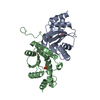
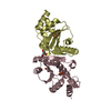
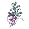
Structure of Phosphopantetheine adenylyltransferase (PPAT) from Enterobacter spp. with the expression tag bound in the substrate binding site of a neighbouring molecule at 2.37 A resolution.
Components
Phosphopantetheine adenylyltransferase
Keywords
TRANSFERASE / coaD / PPAT / COENZYME A biosynthesis
Function / homology
Function and homology information
pantetheine-phosphate adenylyltransferase / pantetheine-phosphate adenylyltransferase activity / coenzyme A biosynthetic process / ATP binding / cytoplasm Similarity search - Function
Journal: To Be Published Title: Structure of Phosphopantetheine adenylyltransferase (PPAT) from Enterobacter spp. with the expression tag bound in the substrate binding site of a neighbouring molecule at 2.37 A resolution. Authors: Ahmad, N. / Sharma, P. / Sharma, S. / Singh, T.P.
 Movie
Movie Controller

 Yorodumi
 Open data
 Basic information
 Components
 Keywords
 Function and homology information
 Enterobacter sp. 638 (bacteria)
 X-RAY DIFFRACTION /
  Authors
 India, 1items
  Citation
 Structure visualization
 Downloads & links
 Other downloads
 PDBj
PDBj Assembly
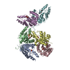

 Mass: 140.032 Da / Num. of mol.: 6 / Source method: obtained synthetically / Formula: C2H5O5P / Feature type: SUBJECT OF INVESTIGATION
 Mass: 92.094 Da / Num. of mol.: 2 / Source method: obtained synthetically / Formula: C3H8O3
 Mass: 62.068 Da / Num. of mol.: 2 / Source method: obtained synthetically / Formula: C2H6O2
 Mass: 18.015 Da / Num. of mol.: 573 / Source method: isolated from a natural source / Formula: H2O
 Sample preparation
 / Beamline: ID23-2 / Wavelength: 0.8731 Å
 Processing