 Movie
Movie Controller


+ Open data

- Basic information
| Entry | Database: PDB / ID: 9ixp | ||||||
|---|---|---|---|---|---|---|---|
| Title | Crystal structure of OXA-10 variant A124T | ||||||
 Components Components | Beta-lactamase OXA-10 | ||||||
 Keywords Keywords | HYDROLASE / Class D Beta-lactamase / OXA-10 variant | ||||||
| Function / homology |  Function and homology information Function and homology informationpenicillin binding / antibiotic catabolic process / cell wall organization / beta-lactamase activity / beta-lactamase / periplasmic space / response to antibiotic / plasma membrane Similarity search - Function | ||||||
| Biological species |   Pseudomonas aeruginosa (bacteria) Pseudomonas aeruginosa (bacteria) | ||||||
| Method |  X-RAY DIFFRACTION / SYNCHROTRON / MOLECULAR REPLACEMENT / Resolution: 2.2 Å X-RAY DIFFRACTION / SYNCHROTRON / MOLECULAR REPLACEMENT / Resolution: 2.2 Å | ||||||
 Authors Authors | Lee, C.E. / Park, Y.S. / Park, H.J. / Kang, L.W. | ||||||
| Funding support |  Korea, Republic Of, 1items Korea, Republic Of, 1items
| ||||||
 Citation Citation | Journal: Emerg Microbes Infect / Year: 2024 Title: Structural insights into alterations in the substrate spectrum of serine-beta-lactamase OXA-10 from Pseudomonas aeruginosa by single amino acid substitutions. Authors: Lee, C.E. / Park, Y. / Park, H. / Kwak, K. / Lee, H. / Yun, J. / Lee, D. / Lee, J.H. / Lee, S.H. / Kang, L.W. | ||||||
| History |
|
- Structure visualization
| Structure viewer | Molecule: MolmilJmol/JSmol |
|---|
- Downloads & links
-Download
| PDBx/mmCIF format | 9ixp.cif.gz | 117.3 KB | Display | PDBx/mmCIF format |
|---|---|---|---|---|
| PDB format | pdb9ixp.ent.gz | 88.7 KB | Display | PDB format |
| PDBx/mmJSON format | 9ixp.json.gz | Tree view | PDBx/mmJSON format | |
| Others |  Other downloads Other downloads |
-Validation report
| Arichive directory | https://data.pdbj.org/pub/pdb/validation_reports/ix/9ixpftp://data.pdbj.org/pub/pdb/validation_reports/ix/9ixp | HTTPS FTP |
|---|
-Related structure data
| Related structure data | 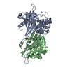 9ixnC  9ixoC  9ixqC  9ixrC  1fofS C: citing same article ( S: Starting model for refinement |
|---|---|
| Similar structure data |
-Links
 PDBj
PDBj- Assembly
| Deposited unit | 
| ||||||||
|---|---|---|---|---|---|---|---|---|---|
| 1 |
| ||||||||
| Unit cell |
|
-Components
| #1: Protein | Mass: 27866.664 Da / Num. of mol.: 2 / Mutation: A124T Source method: isolated from a genetically manipulated source Source: (gene. exp.) Pseudomonas aeruginosa (bacteria) / Gene: bla, oxa10, pse2 / Plasmid: pET-29b / Production host: #2: Water | ChemComp-HOH / |  Mass: 18.015 Da / Num. of mol.: 244 / Source method: isolated from a natural source / Formula: H2O Mass: 18.015 Da / Num. of mol.: 244 / Source method: isolated from a natural source / Formula: H2OHas ligand of interest | Y | Has protein modification | Y | |
|---|
-Experimental details
-Experiment
| Experiment | Method: X-RAY DIFFRACTION / Number of used crystals: 1 |
|---|
- Sample preparation
| Crystal | Density Matthews: 2.86 Å3/Da / Density % sol: 56.93 % |
|---|---|
| Crystal grow | Temperature: 288 K / Method: vapor diffusion, hanging drop Details: 20% PEG 3350, 0.2 M Sodium formate, 0.1 M Tris-HCl pH 7.0 |
-Data collection
| Diffraction | Mean temperature: 100 K / Ambient temp details: Steady flow of liquid nitrogen / Serial crystal experiment: N |
|---|---|
| Diffraction source | Source: SYNCHROTRON / Site: PAL/PLS / Beamline: 11C / Wavelength: 0.97942 Å |
| Detector | Type: DECTRIS PILATUS 6M / Detector: PIXEL / Date: Dec 5, 2023 |
| Radiation | Protocol: SINGLE WAVELENGTH / Monochromatic (M) / Laue (L): M / Scattering type: x-ray |
| Radiation wavelength | Wavelength: 0.97942 Å / Relative weight: 1 |
| Reflection | Resolution: 2.2→47.53 Å / Num. obs: 31969 / % possible obs: 96.15 % / Redundancy: 6.3 % / CC1/2: 0.996 / CC star: 0.999 / Rmerge(I) obs: 0.145 / Rpim(I) all: 0.056 / Rrim(I) all: 0.156 / Rsym value: 0.153 / Net I/σ(I): 11.39 |
| Reflection shell | Resolution: 2.2→2.28 Å / Num. unique obs: 3033 / Rpim(I) all: 0.26 |
- Processing
| Software |
| ||||||||||||||||||||||||||||||||||||||||||||||||||||||||||||||||||||||||||||||||||||||||||||||||||||||||||||||||||||||||||||||||||||||||||||||||||||||||||||||||||||||||||||||||||||||
|---|---|---|---|---|---|---|---|---|---|---|---|---|---|---|---|---|---|---|---|---|---|---|---|---|---|---|---|---|---|---|---|---|---|---|---|---|---|---|---|---|---|---|---|---|---|---|---|---|---|---|---|---|---|---|---|---|---|---|---|---|---|---|---|---|---|---|---|---|---|---|---|---|---|---|---|---|---|---|---|---|---|---|---|---|---|---|---|---|---|---|---|---|---|---|---|---|---|---|---|---|---|---|---|---|---|---|---|---|---|---|---|---|---|---|---|---|---|---|---|---|---|---|---|---|---|---|---|---|---|---|---|---|---|---|---|---|---|---|---|---|---|---|---|---|---|---|---|---|---|---|---|---|---|---|---|---|---|---|---|---|---|---|---|---|---|---|---|---|---|---|---|---|---|---|---|---|---|---|---|---|---|---|---|
| Refinement | Method to determine structure: MOLECULAR REPLACEMENT Starting model: 1FOF Resolution: 2.2→47.53 Å / Cor.coef. Fo:Fc: 0.956 / Cor.coef. Fo:Fc free: 0.932 / SU B: 7.531 / SU ML: 0.179 / Cross valid method: THROUGHOUT / ESU R: 0.243 / ESU R Free: 0.204 / Stereochemistry target values: MAXIMUM LIKELIHOOD / Details: HYDROGENS HAVE BEEN ADDED IN THE RIDING POSITIONS
| ||||||||||||||||||||||||||||||||||||||||||||||||||||||||||||||||||||||||||||||||||||||||||||||||||||||||||||||||||||||||||||||||||||||||||||||||||||||||||||||||||||||||||||||||||||||
| Solvent computation | Ion probe radii: 0.8 Å / Shrinkage radii: 0.8 Å / VDW probe radii: 1.2 Å / Solvent model: MASK | ||||||||||||||||||||||||||||||||||||||||||||||||||||||||||||||||||||||||||||||||||||||||||||||||||||||||||||||||||||||||||||||||||||||||||||||||||||||||||||||||||||||||||||||||||||||
| Displacement parameters | Biso mean: 42.955 Å2
| ||||||||||||||||||||||||||||||||||||||||||||||||||||||||||||||||||||||||||||||||||||||||||||||||||||||||||||||||||||||||||||||||||||||||||||||||||||||||||||||||||||||||||||||||||||||
| Refinement step | Cycle: 1 / Resolution: 2.2→47.53 Å
| ||||||||||||||||||||||||||||||||||||||||||||||||||||||||||||||||||||||||||||||||||||||||||||||||||||||||||||||||||||||||||||||||||||||||||||||||||||||||||||||||||||||||||||||||||||||
| Refine LS restraints |
|