[English] 日本語
 Yorodumi
- PDB-9iv9: Cryo-EM structure of a truncated Nipah Virus L Protein bound by P... -
+ Open data
- Basic information
| Entry | Database: PDB / ID: 9iv9 | |||||||||||||||||||||||||||
|---|---|---|---|---|---|---|---|---|---|---|---|---|---|---|---|---|---|---|---|---|---|---|---|---|---|---|---|---|
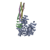
| Title | Cryo-EM structure of a truncated Nipah Virus L Protein bound by Phosphoprotein Tetramer | |||||||||||||||||||||||||||
 Components Components |
| |||||||||||||||||||||||||||
 Keywords Keywords | VIRAL PROTEIN / RNA polymerase | |||||||||||||||||||||||||||
| Function / homology |  Function and homology information Function and homology informationnegative stranded viral RNA transcription / NNS virus cap methyltransferase / GDP polyribonucleotidyltransferase / negative stranded viral RNA replication / Hydrolases; Acting on acid anhydrides; In phosphorus-containing anhydrides / virion component / molecular adaptor activity / host cell cytoplasm / mRNA 5'-cap (guanine-N7-)-methyltransferase activity / symbiont-mediated suppression of host innate immune response ...negative stranded viral RNA transcription / NNS virus cap methyltransferase / GDP polyribonucleotidyltransferase / negative stranded viral RNA replication / Hydrolases; Acting on acid anhydrides; In phosphorus-containing anhydrides / virion component / molecular adaptor activity / host cell cytoplasm / mRNA 5'-cap (guanine-N7-)-methyltransferase activity / symbiont-mediated suppression of host innate immune response / RNA-directed RNA polymerase / RNA-directed RNA polymerase activity / GTPase activity / ATP binding Similarity search - Function | |||||||||||||||||||||||||||
| Biological species |  Henipavirus nipahense Henipavirus nipahense | |||||||||||||||||||||||||||
| Method | ELECTRON MICROSCOPY / single particle reconstruction / cryo EM / Resolution: 2.31 Å | |||||||||||||||||||||||||||
 Authors Authors | Xue, L. / Chang, T. / Gui, J. / Li, Z. / Zhao, H. / Zou, B. / Li, M. / He, J. / Chen, X. / Xiong, X. | |||||||||||||||||||||||||||
| Funding support |  China, 1items China, 1items
| |||||||||||||||||||||||||||
 Citation Citation |  Journal: Protein Cell / Year: 2025 Journal: Protein Cell / Year: 2025Title: Cryo-EM structures of Nipah virus polymerase complex reveal highly varied interactions between L and P proteins among paramyxoviruses. Authors: Lu Xue / Tiancai Chang / Jiacheng Gui / Zimu Li / Heyu Zhao / Binqian Zou / Junnan Lu / Mei Li / Xin Wen / Shenghua Gao / Peng Zhan / Lijun Rong / Liqiang Feng / Peng Gong / Jun He / Xinwen ...Authors: Lu Xue / Tiancai Chang / Jiacheng Gui / Zimu Li / Heyu Zhao / Binqian Zou / Junnan Lu / Mei Li / Xin Wen / Shenghua Gao / Peng Zhan / Lijun Rong / Liqiang Feng / Peng Gong / Jun He / Xinwen Chen / Xiaoli Xiong /   Abstract: Nipah virus (NiV) and related viruses form a distinct henipavirus genus within the Paramyxoviridae family. NiV continues to spillover into the humans causing deadly outbreaks with increasing human- ...Nipah virus (NiV) and related viruses form a distinct henipavirus genus within the Paramyxoviridae family. NiV continues to spillover into the humans causing deadly outbreaks with increasing human-bat interaction. NiV encodes the large protein (L) and phosphoprotein (P) to form the viral RNA polymerase machinery. Their sequences show limited homologies to those of non-henipavirus paramyxoviruses. We report two cryo-electron microscopy (cryo-EM) structures of the Nipah virus (NiV) polymerase L-P complex, expressed and purified in either its full-length or truncated form. The structures resolve the RNA-dependent RNA polymerase (RdRp) and polyribonucleotidyl transferase (PRNTase) domains of the L protein, as well as a tetrameric P protein bundle bound to the L-RdRp domain. L-protein C-terminal regions are unresolved, indicating flexibility. Two PRNTase domain zinc-binding sites, conserved in most Mononegavirales, are confirmed essential for NiV polymerase activity. The structures further reveal anchoring of the P protein bundle and P protein X domain (XD) linkers on L, via an interaction pattern distinct among Paramyxoviridae. These interactions facilitate binding of a P protein XD linker in the nucleotide entry channel and distinct positioning of other XD linkers. We show that the disruption of the L-P interactions reduces NiV polymerase activity. The reported structures should facilitate rational antiviral-drug discovery and provide a guide for the functional study of NiV polymerase. | |||||||||||||||||||||||||||
| History |
|
- Structure visualization
| Structure viewer | Molecule:  Molmil Molmil Jmol/JSmol Jmol/JSmol |
|---|
- Downloads & links
- Download
| PDBx/mmCIF format |  9iv9.cif.gz 9iv9.cif.gz | 371.3 KB | Display |  PDBx/mmCIF format PDBx/mmCIF format |
|---|---|---|---|---|
| PDB format |  pdb9iv9.ent.gz pdb9iv9.ent.gz | 275.2 KB | Display |  PDB format PDB format |
| PDBx/mmJSON format |  9iv9.json.gz 9iv9.json.gz | Tree view |  PDBx/mmJSON format PDBx/mmJSON format | |
| Others |  Other downloads Other downloads |
-Validation report
| Arichive directory |  https://data.pdbj.org/pub/pdb/validation_reports/iv/9iv9 https://data.pdbj.org/pub/pdb/validation_reports/iv/9iv9 ftp://data.pdbj.org/pub/pdb/validation_reports/iv/9iv9 ftp://data.pdbj.org/pub/pdb/validation_reports/iv/9iv9 | HTTPS FTP |
|---|
-Related structure data
| Related structure data |  60922MC  9ivaC M: map data used to model this data C: citing same article ( |
|---|---|
| Similar structure data | Similarity search - Function & homology  F&H Search F&H Search |
- Links
Links
- Assembly
| Deposited unit | 
|
|---|---|
| 1 |
|
- Components
| #1: Protein | Mass: 166266.891 Da / Num. of mol.: 1 Source method: isolated from a genetically manipulated source Source: (gene. exp.)  Henipavirus nipahense / Production host: Henipavirus nipahense / Production host:  Spodoptera frugiperda ascovirus 1c Spodoptera frugiperda ascovirus 1cReferences: UniProt: Q997F0, RNA-directed RNA polymerase, Hydrolases; Acting on acid anhydrides; In phosphorus-containing anhydrides, GDP polyribonucleotidyltransferase, NNS virus cap methyltransferase | ||||||
|---|---|---|---|---|---|---|---|
| #2: Protein | Mass: 78390.320 Da / Num. of mol.: 4 Source method: isolated from a genetically manipulated source Source: (gene. exp.)  Henipavirus nipahense / Gene: P/V/C / Production host: Henipavirus nipahense / Gene: P/V/C / Production host:  #3: Chemical | Has ligand of interest | Y | Has protein modification | N | |
-Experimental details
-Experiment
| Experiment | Method: ELECTRON MICROSCOPY |
|---|---|
| EM experiment | Aggregation state: PARTICLE / 3D reconstruction method: single particle reconstruction |
- Sample preparation
| Component | Name: Cryo-EM structure of a truncated Nipah Virus L Protein bound by Phosphoprotein Tetramer Type: COMPLEX / Entity ID: #1-#2 / Source: RECOMBINANT |
|---|---|
| Source (natural) | Organism:  Henipavirus nipahense Henipavirus nipahense |
| Source (recombinant) | Organism:  |
| Buffer solution | pH: 8 |
| Specimen | Embedding applied: NO / Shadowing applied: NO / Staining applied: NO / Vitrification applied: YES |
| Vitrification | Cryogen name: ETHANE |
- Electron microscopy imaging
| Experimental equipment |  Model: Titan Krios / Image courtesy: FEI Company |
|---|---|
| Microscopy | Model: TFS KRIOS |
| Electron gun | Electron source:  FIELD EMISSION GUN / Accelerating voltage: 300 kV / Illumination mode: FLOOD BEAM FIELD EMISSION GUN / Accelerating voltage: 300 kV / Illumination mode: FLOOD BEAM |
| Electron lens | Mode: BRIGHT FIELD / Nominal defocus max: 2400 nm / Nominal defocus min: 600 nm |
| Image recording | Electron dose: 50 e/Å2 / Film or detector model: TFS FALCON 4i (4k x 4k) |
- Processing
| EM software | Name: PHENIX / Category: model refinement | ||||||||||||||||||||||||
|---|---|---|---|---|---|---|---|---|---|---|---|---|---|---|---|---|---|---|---|---|---|---|---|---|---|
| CTF correction | Type: PHASE FLIPPING AND AMPLITUDE CORRECTION | ||||||||||||||||||||||||
| 3D reconstruction | Resolution: 2.31 Å / Resolution method: FSC 0.143 CUT-OFF / Num. of particles: 674945 / Symmetry type: POINT | ||||||||||||||||||||||||
| Refinement | Highest resolution: 2.31 Å Stereochemistry target values: REAL-SPACE (WEIGHTED MAP SUM AT ATOM CENTERS) | ||||||||||||||||||||||||
| Refine LS restraints |
|
 Movie
Movie Controller


 PDBj
PDBj


