 Movie
Movie Controller

[English] 日本語
 Yorodumi
- PDB-9iud: High resolution structure of Lectin-Like ox-LDL Receptor 1 with B... -
+ Open data

- Basic information
| Entry | Database: PDB / ID: 9iud | ||||||
|---|---|---|---|---|---|---|---|
| Title | High resolution structure of Lectin-Like ox-LDL Receptor 1 with BI-0115 in space group P 21 21 21 | ||||||
 Components Components | Oxidized low-density lipoprotein receptor 1 | ||||||
 Keywords Keywords | LIPID BINDING PROTEIN / inhibitor / complex | ||||||
| Function / homology |  Function and homology information Function and homology informationlow-density lipoprotein particle receptor activity / lipoprotein metabolic process / leukocyte cell-cell adhesion / blood circulation / tertiary granule membrane / immune system process / specific granule membrane / Cell surface interactions at the vascular wall / carbohydrate binding / signaling receptor complex ...low-density lipoprotein particle receptor activity / lipoprotein metabolic process / leukocyte cell-cell adhesion / blood circulation / tertiary granule membrane / immune system process / specific granule membrane / Cell surface interactions at the vascular wall / carbohydrate binding / signaling receptor complex / membrane raft / inflammatory response / Neutrophil degranulation / proteolysis / extracellular region / nucleoplasm / membrane / identical protein binding / plasma membrane Similarity search - Function | ||||||
| Biological species |  Homo sapiens (human) Homo sapiens (human) | ||||||
| Method |  X-RAY DIFFRACTION / SYNCHROTRON / MOLECULAR REPLACEMENT / Resolution: 1.98 Å X-RAY DIFFRACTION / SYNCHROTRON / MOLECULAR REPLACEMENT / Resolution: 1.98 Å | ||||||
 Authors Authors | Khan, M.A. / Arulandu, A. | ||||||
| Funding support |  India, 1items India, 1items
| ||||||
 Citation Citation | Journal: To Be Published Title: High resolution structure of Lectin-Like ox-LDL Receptor 1 with BI-0115 in space group P 21 21 21 Authors: khan, M.A. / Arulandu, A. | ||||||
| History |
|
- Structure visualization
| Structure viewer | Molecule: MolmilJmol/JSmol |
|---|
- Downloads & links
-Download
| PDBx/mmCIF format | 9iud.cif.gz | 234.9 KB | Display | PDBx/mmCIF format |
|---|---|---|---|---|
| PDB format | pdb9iud.ent.gz | Display | PDB format | |
| PDBx/mmJSON format | 9iud.json.gz | Tree view | PDBx/mmJSON format | |
| Others |  Other downloads Other downloads |
-Validation report
| Arichive directory | https://data.pdbj.org/pub/pdb/validation_reports/iu/9iudftp://data.pdbj.org/pub/pdb/validation_reports/iu/9iud | HTTPS FTP |
|---|
-Related structure data
| Similar structure data |
|---|
-Links
 PDBj
PDBj


- Assembly
| Deposited unit | 
| ||||||||
|---|---|---|---|---|---|---|---|---|---|
| 1 | 
| ||||||||
| 2 | 
| ||||||||
| Unit cell |
|
-Components
| #1: Protein | Mass: 15668.785 Da / Num. of mol.: 8 Source method: isolated from a genetically manipulated source Source: (gene. exp.) Homo sapiens (human) / Gene: OLR1, CLEC8A, LOX1 / Production host:  #2: Chemical | ChemComp-NJT / 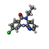  Mass: 287.744 Da / Num. of mol.: 4 / Source method: obtained synthetically / Formula: C15H14ClN3O / Feature type: SUBJECT OF INVESTIGATION Mass: 287.744 Da / Num. of mol.: 4 / Source method: obtained synthetically / Formula: C15H14ClN3O / Feature type: SUBJECT OF INVESTIGATION#3: Chemical | ChemComp-GOL /   Mass: 92.094 Da / Num. of mol.: 9 / Source method: obtained synthetically / Formula: C3H8O3 Mass: 92.094 Da / Num. of mol.: 9 / Source method: obtained synthetically / Formula: C3H8O3#4: Water | ChemComp-HOH / |  Mass: 18.015 Da / Num. of mol.: 699 / Source method: isolated from a natural source / Formula: H2O Mass: 18.015 Da / Num. of mol.: 699 / Source method: isolated from a natural source / Formula: H2OHas ligand of interest | Y | Has protein modification | Y | |
|---|
-Experimental details
-Experiment
| Experiment | Method: X-RAY DIFFRACTION / Number of used crystals: 1 |
|---|
- Sample preparation
| Crystal | Density Matthews: 2.22 Å3/Da / Density % sol: 44.6 % |
|---|---|
| Crystal grow | Temperature: 293.15 K / Method: vapor diffusion, sitting drop Details: 0.2 M Magnesium formate dihydrate, 20 % w/v PEG 3350 |
-Data collection
| Diffraction | Mean temperature: 100 K / Serial crystal experiment: N | ||||||||||||||||||||||||
|---|---|---|---|---|---|---|---|---|---|---|---|---|---|---|---|---|---|---|---|---|---|---|---|---|---|
| Diffraction source | Source: SYNCHROTRON / Site: ELETTRA  / Beamline: 11.2C / Wavelength: 1 Å / Beamline: 11.2C / Wavelength: 1 Å | ||||||||||||||||||||||||
| Detector | Type: DECTRIS PILATUS 6M / Detector: PIXEL / Date: May 12, 2022 | ||||||||||||||||||||||||
| Radiation | Protocol: SINGLE WAVELENGTH / Monochromatic (M) / Laue (L): M / Scattering type: x-ray | ||||||||||||||||||||||||
| Radiation wavelength | Wavelength: 1 Å / Relative weight: 1 | ||||||||||||||||||||||||
| Reflection | Resolution: 1.97→80.25 Å / Num. obs: 71550 / % possible obs: 94.9 % / Redundancy: 9.6 % / CC1/2: 0.997 / Rmerge(I) obs: 0.121 / Rpim(I) all: 0.041 / Rrim(I) all: 0.128 / Net I/σ(I): 11.3 | ||||||||||||||||||||||||
| Reflection shell | Diffraction-ID: 1
|
- Processing
| Software |
| |||||||||||||||||||||||||||||||||||||||||||||||||||||||||||||||||||||||||||||||||||||||||||||||||||||||||||||||||||||||||||||||||||||||||||||||||||||||||||||||||||||||||||||||||||||||||||||
|---|---|---|---|---|---|---|---|---|---|---|---|---|---|---|---|---|---|---|---|---|---|---|---|---|---|---|---|---|---|---|---|---|---|---|---|---|---|---|---|---|---|---|---|---|---|---|---|---|---|---|---|---|---|---|---|---|---|---|---|---|---|---|---|---|---|---|---|---|---|---|---|---|---|---|---|---|---|---|---|---|---|---|---|---|---|---|---|---|---|---|---|---|---|---|---|---|---|---|---|---|---|---|---|---|---|---|---|---|---|---|---|---|---|---|---|---|---|---|---|---|---|---|---|---|---|---|---|---|---|---|---|---|---|---|---|---|---|---|---|---|---|---|---|---|---|---|---|---|---|---|---|---|---|---|---|---|---|---|---|---|---|---|---|---|---|---|---|---|---|---|---|---|---|---|---|---|---|---|---|---|---|---|---|---|---|---|---|---|---|---|
| Refinement | Method to determine structure: MOLECULAR REPLACEMENT / Resolution: 1.98→41.27 Å / SU ML: 0.24 / Cross valid method: THROUGHOUT / σ(F): 1.34 / Phase error: 29.74 / Stereochemistry target values: ML
| |||||||||||||||||||||||||||||||||||||||||||||||||||||||||||||||||||||||||||||||||||||||||||||||||||||||||||||||||||||||||||||||||||||||||||||||||||||||||||||||||||||||||||||||||||||||||||||
| Solvent computation | Ion probe radii: 0.7 Å / Shrinkage radii: 0.9 Å / VDW probe radii: 1.11 Å / Solvent model: FLAT BULK SOLVENT MODEL | |||||||||||||||||||||||||||||||||||||||||||||||||||||||||||||||||||||||||||||||||||||||||||||||||||||||||||||||||||||||||||||||||||||||||||||||||||||||||||||||||||||||||||||||||||||||||||||
| Displacement parameters | Biso mean: 33 Å2 | |||||||||||||||||||||||||||||||||||||||||||||||||||||||||||||||||||||||||||||||||||||||||||||||||||||||||||||||||||||||||||||||||||||||||||||||||||||||||||||||||||||||||||||||||||||||||||||
| Refinement step | Cycle: LAST / Resolution: 1.98→41.27 Å
| |||||||||||||||||||||||||||||||||||||||||||||||||||||||||||||||||||||||||||||||||||||||||||||||||||||||||||||||||||||||||||||||||||||||||||||||||||||||||||||||||||||||||||||||||||||||||||||
| Refine LS restraints |
| |||||||||||||||||||||||||||||||||||||||||||||||||||||||||||||||||||||||||||||||||||||||||||||||||||||||||||||||||||||||||||||||||||||||||||||||||||||||||||||||||||||||||||||||||||||||||||||
| LS refinement shell |
|
