 Movie
Movie Controller

[English] 日本語
 Yorodumi
- PDB-9iqj: Structure of oleate hydratase mutant L151V from Staphylococcus au... -
+ Open data

- Basic information
| Entry | Database: PDB / ID: 9iqj | ||||||
|---|---|---|---|---|---|---|---|
| Title | Structure of oleate hydratase mutant L151V from Staphylococcus aureus in the complex with linoleic acid | ||||||
 Components Components | Oleate hydratase | ||||||
 Keywords Keywords | LYASE / Oleate Hydratase | ||||||
| Function / homology |  Function and homology information Function and homology informationoleate hydratase / oleate hydratase activity / FAD binding / fatty acid metabolic process Similarity search - Function | ||||||
| Biological species |   Staphylococcus aureus (bacteria) Staphylococcus aureus (bacteria) | ||||||
| Method |  X-RAY DIFFRACTION / SYNCHROTRON / MOLECULAR REPLACEMENT / Resolution: 1.64 Å X-RAY DIFFRACTION / SYNCHROTRON / MOLECULAR REPLACEMENT / Resolution: 1.64 Å | ||||||
 Authors Authors | Xue, S. / Feng, T. | ||||||
| Funding support |  China, 1items China, 1items
| ||||||
 Citation Citation | Journal: To Be Published Title: Cooperative Evolution of Oleate Hydratase via Combinatorial Distal Site Mutations Authors: Xue, S. / Feng, T. | ||||||
| History |
|
- Structure visualization
| Structure viewer | Molecule: MolmilJmol/JSmol |
|---|
- Downloads & links
-Download
| PDBx/mmCIF format | 9iqj.cif.gz | 500.8 KB | Display | PDBx/mmCIF format |
|---|---|---|---|---|
| PDB format | pdb9iqj.ent.gz | 325.9 KB | Display | PDB format |
| PDBx/mmJSON format | 9iqj.json.gz | Tree view | PDBx/mmJSON format | |
| Others |  Other downloads Other downloads |
-Validation report
| Summary document | 9iqj_validation.pdf.gz | 970.5 KB | Display | wwPDB validaton report |
|---|---|---|---|---|
| Full document | 9iqj_full_validation.pdf.gz | 982.2 KB | Display | |
| Data in XML | 9iqj_validation.xml.gz | 98.6 KB | Display | |
| Data in CIF | 9iqj_validation.cif.gz | 142.4 KB | Display | |
| Arichive directory | https://data.pdbj.org/pub/pdb/validation_reports/iq/9iqjftp://data.pdbj.org/pub/pdb/validation_reports/iq/9iqj | HTTPS FTP |
-Related structure data
| Related structure data |  9iqaC  9iqiC 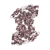 7kawS C: citing same article ( S: Starting model for refinement |
|---|---|
| Similar structure data |
-Links
 PDBj
PDBj- Assembly
| Deposited unit | 
| ||||||||||||
|---|---|---|---|---|---|---|---|---|---|---|---|---|---|
| 1 | 
| ||||||||||||
| 2 | 
| ||||||||||||
| Unit cell |
| ||||||||||||
| Components on special symmetry positions |
|
-Components
| #1: Protein | Mass: 67845.484 Da / Num. of mol.: 3 / Mutation: L151V Source method: isolated from a genetically manipulated source Source: (gene. exp.) Staphylococcus aureus (bacteria)Gene: EP54_06595, EQ90_12415, GO814_02765, GO942_14045, GZ163_10760, HMPREF3211_02399 Production host: #2: Chemical |   Mass: 280.445 Da / Num. of mol.: 3 / Source method: obtained synthetically / Formula: C18H32O2 / Feature type: SUBJECT OF INVESTIGATION Mass: 280.445 Da / Num. of mol.: 3 / Source method: obtained synthetically / Formula: C18H32O2 / Feature type: SUBJECT OF INVESTIGATION#3: Chemical | ChemComp-PEG /   Mass: 106.120 Da / Num. of mol.: 4 / Source method: obtained synthetically / Formula: C4H10O3 Mass: 106.120 Da / Num. of mol.: 4 / Source method: obtained synthetically / Formula: C4H10O3#4: Chemical | ChemComp-GOL / |   Mass: 92.094 Da / Num. of mol.: 1 / Source method: obtained synthetically / Formula: C3H8O3 Mass: 92.094 Da / Num. of mol.: 1 / Source method: obtained synthetically / Formula: C3H8O3#5: Water | ChemComp-HOH / |  Mass: 18.015 Da / Num. of mol.: 2251 / Source method: isolated from a natural source / Formula: H2O Mass: 18.015 Da / Num. of mol.: 2251 / Source method: isolated from a natural source / Formula: H2OHas ligand of interest | Y | Has protein modification | N | |
|---|
-Experimental details
-Experiment
| Experiment | Method: X-RAY DIFFRACTION / Number of used crystals: 1 |
|---|
- Sample preparation
| Crystal | Density Matthews: 2.82 Å3/Da / Density % sol: 56.32 % |
|---|---|
| Crystal grow | Temperature: 300 K / Method: vapor diffusion, hanging drop Details: 0.2 M Ammooium citrate tribasic pH 7.0, 15 % w/v Polyethylene glycol 3350 |
-Data collection
| Diffraction | Mean temperature: 100 K / Serial crystal experiment: N |
|---|---|
| Diffraction source | Source: SYNCHROTRON / Site: SSRF / Beamline: BL17B1 / Wavelength: 0.9793 Å |
| Detector | Type: MAR CCD 165 mm / Detector: CCD / Date: Jan 9, 2024 |
| Radiation | Protocol: SINGLE WAVELENGTH / Monochromatic (M) / Laue (L): M / Scattering type: x-ray |
| Radiation wavelength | Wavelength: 0.9793 Å / Relative weight: 1 |
| Reflection | Resolution: 1.64→37.82 Å / Num. obs: 274186 / % possible obs: 99.34 % / Redundancy: 6.8 % / Biso Wilson estimate: 14.7 Å2 / CC1/2: 0.993 / CC star: 0.998 / Rmerge(I) obs: 0.1108 / Rrim(I) all: 0.1201 / Net I/av σ(I): 14.58 / Net I/σ(I): 3.66 |
| Reflection shell | Resolution: 1.64→1.69 Å / Redundancy: 6.8 % / Rmerge(I) obs: 0.4172 / Mean I/σ(I) obs: 3.66 / Num. unique obs: 26636 / CC1/2: 0.949 / CC star: 0.987 / Rrim(I) all: 0.4509 / % possible all: 96.94 |
- Processing
| Software |
| |||||||||||||||||||||||||||||||||||||||||||||||||||||||||||||||||||||||||||||||||||||||||||||||||||||||||
|---|---|---|---|---|---|---|---|---|---|---|---|---|---|---|---|---|---|---|---|---|---|---|---|---|---|---|---|---|---|---|---|---|---|---|---|---|---|---|---|---|---|---|---|---|---|---|---|---|---|---|---|---|---|---|---|---|---|---|---|---|---|---|---|---|---|---|---|---|---|---|---|---|---|---|---|---|---|---|---|---|---|---|---|---|---|---|---|---|---|---|---|---|---|---|---|---|---|---|---|---|---|---|---|---|---|---|
| Refinement | Method to determine structure: MOLECULAR REPLACEMENT Starting model: 7KAW Resolution: 1.64→37.82 Å / SU ML: 0.1338 / Cross valid method: FREE R-VALUE / σ(F): 1.37 / Phase error: 17.3349 Stereochemistry target values: GeoStd + Monomer Library + CDL v1.2
| |||||||||||||||||||||||||||||||||||||||||||||||||||||||||||||||||||||||||||||||||||||||||||||||||||||||||
| Solvent computation | Shrinkage radii: 0.9 Å / VDW probe radii: 1.1 Å / Solvent model: FLAT BULK SOLVENT MODEL | |||||||||||||||||||||||||||||||||||||||||||||||||||||||||||||||||||||||||||||||||||||||||||||||||||||||||
| Displacement parameters | Biso mean: 19.84 Å2 | |||||||||||||||||||||||||||||||||||||||||||||||||||||||||||||||||||||||||||||||||||||||||||||||||||||||||
| Refinement step | Cycle: LAST / Resolution: 1.64→37.82 Å
| |||||||||||||||||||||||||||||||||||||||||||||||||||||||||||||||||||||||||||||||||||||||||||||||||||||||||
| Refine LS restraints |
| |||||||||||||||||||||||||||||||||||||||||||||||||||||||||||||||||||||||||||||||||||||||||||||||||||||||||
| LS refinement shell |
|
