| 登録情報 | データベース: PDB / ID: 9int
|
|---|
| タイトル | Crystal structure of the complex of the beta,kappa-carrageenase Cgbk16A from Wenyingzhuangia fucanilytica with an oligosaccharide of furcellaran |
|---|
 要素 要素 | GH16 domain-containing protein |
|---|
 キーワード キーワード | HYDROLASE / complex / Carrageenase / GH16_13 / Glucoside hydrolase / oligotetrasaccharide / furcellaran |
|---|
| 機能・相同性 | : / Glycosyl hydrolases family 16 / Glycoside hydrolase family 16 / Glycosyl hydrolases family 16 (GH16) domain profile. / hydrolase activity, hydrolyzing O-glycosyl compounds / Concanavalin A-like lectin/glucanase domain superfamily / carbohydrate metabolic process / GH16 domain-containing protein 機能・相同性情報 機能・相同性情報 |
|---|
| 生物種 |  Wenyingzhuangia fucanilytica (バクテリア) Wenyingzhuangia fucanilytica (バクテリア) |
|---|
| 手法 |  X線回折 / シンクロトロン / 分子置換 / 解像度: 1.56 Å X線回折 / シンクロトロン / 分子置換 / 解像度: 1.56 Å |
|---|
 データ登録者 データ登録者 | Chang, Y. / Chen, F. |
|---|
| 資金援助 | 1件 | 組織 | 認可番号 | 国 |
|---|
| Other government | 2023YFD2100605 | |
|
|---|
 引用 引用 | ジャーナル: J.Agric.Food Chem. / 年: 2024
タイトル: Structural Insights into the Substrate Recognition and Catalytic Mechanism of a GH16 beta kappa-Carrageenase from Wenyingzhuangia fucanilytica.
著者: Chen, F. / Xue, C. / Chen, G. / Mei, X. / Zheng, L. / Chang, Y. |
|---|
| 履歴 | | 登録 | 2024年7月8日 | 登録サイト: PDBJ / 処理サイト: PDBC |
|---|
| 改定 1.0 | 2024年7月17日 | Provider: repository / タイプ: Initial release |
|---|
| 改定 1.1 | 2024年7月31日 | Group: Database references / Structure summary / カテゴリ: citation / struct / Item: _citation.title / _struct.title |
|---|
| 改定 1.2 | 2024年9月25日 | Group: Database references / カテゴリ: citation / citation_author
Item: _citation.country / _citation.journal_abbrev ..._citation.country / _citation.journal_abbrev / _citation.journal_id_CSD / _citation.journal_id_ISSN / _citation.journal_volume / _citation.page_first / _citation.page_last / _citation.pdbx_database_id_DOI / _citation.pdbx_database_id_PubMed / _citation.title / _citation.year |
|---|
|
|---|
 ムービー
ムービー コントローラー


 データを開く
 基本情報
基本情報 構造の表示
構造の表示 ダウンロードとリンク
 その他のダウンロード
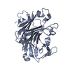
 PDBj
PDBj
 集合体
集合体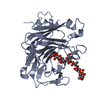
 分子量: 18.015 Da / 分子数: 437 / 由来タイプ: 天然 / 式: H2O
 試料調製
試料調製 / ビームライン: BL19U1 / 波長: 0.987 Å
 解析
解析