 Movie
Movie Controller

[English] 日本語
 Yorodumi
- PDB-9ilt: Crystal structure of alternative complex III from Chloroflexus au... -
+ Open data

- Basic information
| Entry | Database: PDB / ID: 9ilt | ||||||
|---|---|---|---|---|---|---|---|
| Title | Crystal structure of alternative complex III from Chloroflexus aurantiacus | ||||||
 Components Components |
| ||||||
 Keywords Keywords | MEMBRANE PROTEIN / alternative complex III / Chloroflexus aurantiacus / photosynthetic electron transport chains / quinol: electron acceptor oxidoreductase | ||||||
| Function / homology |  Function and homology information Function and homology informationelectron transfer activity / oxidoreductase activity / heme binding / metal ion binding / membrane / plasma membrane Similarity search - Function | ||||||
| Biological species |   Chloroflexus aurantiacus J-10-fl (bacteria) Chloroflexus aurantiacus J-10-fl (bacteria) | ||||||
| Method |  X-RAY DIFFRACTION / SYNCHROTRON / MOLECULAR REPLACEMENT / Resolution: 3.25 Å X-RAY DIFFRACTION / SYNCHROTRON / MOLECULAR REPLACEMENT / Resolution: 3.25 Å | ||||||
 Authors Authors | Xu, X. / Wu, W. | ||||||
| Funding support |  China, 1items China, 1items
| ||||||
 Citation Citation | Journal: Structure / Year: 2025 Title: Crystal structure of the alternative complex III from the phototrophic bacterium Chloroflexus aurantiacus. Authors: Wu, W. / Fang, H. / He, H. / Wu, J. / Gong, Z. / Li, C. / Pei, X. / Xu, X. | ||||||
| History |
|
- Structure visualization
| Structure viewer | Molecule: MolmilJmol/JSmol |
|---|
- Downloads & links
-Download
| PDBx/mmCIF format | 9ilt.cif.gz | 1006.3 KB | Display | PDBx/mmCIF format |
|---|---|---|---|---|
| PDB format | pdb9ilt.ent.gz | 829.4 KB | Display | PDB format |
| PDBx/mmJSON format | 9ilt.json.gz | Tree view | PDBx/mmJSON format | |
| Others |  Other downloads Other downloads |
-Validation report
| Summary document | 9ilt_validation.pdf.gz | 2.3 MB | Display | wwPDB validaton report |
|---|---|---|---|---|
| Full document | 9ilt_full_validation.pdf.gz | 2.3 MB | Display | |
| Data in XML | 9ilt_validation.xml.gz | 93.7 KB | Display | |
| Data in CIF | 9ilt_validation.cif.gz | 117.2 KB | Display | |
| Arichive directory | https://data.pdbj.org/pub/pdb/validation_reports/il/9iltftp://data.pdbj.org/pub/pdb/validation_reports/il/9ilt | HTTPS FTP |
-Related structure data
| Related structure data | 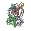 8k9fS S: Starting model for refinement |
|---|---|
| Similar structure data |
-Links
 PDBj
PDBj


- Assembly
| Deposited unit | 
| ||||||||
|---|---|---|---|---|---|---|---|---|---|
| 1 |
| ||||||||
| Unit cell |
|
-Components
-Protein , 5 types, 5 molecules ABCEG
| #1: Protein | Mass: 25247.039 Da / Num. of mol.: 1 / Source method: isolated from a natural source Source: (natural) Chloroflexus aurantiacus J-10-fl (bacteria)References: UniProt: A9WEV2 |
|---|---|
| #2: Protein | Mass: 113115.359 Da / Num. of mol.: 1 / Source method: isolated from a natural source Source: (natural) Chloroflexus aurantiacus J-10-fl (bacteria)References: UniProt: A9WEV3 |
| #3: Protein | Mass: 55223.520 Da / Num. of mol.: 1 / Source method: isolated from a natural source Source: (natural) Chloroflexus aurantiacus J-10-fl (bacteria)References: UniProt: A9WEV4 |
| #5: Protein | Mass: 23017.016 Da / Num. of mol.: 1 / Source method: isolated from a natural source Source: (natural) Chloroflexus aurantiacus J-10-fl (bacteria)References: UniProt: A9WEV6 |
| #7: Protein | Mass: 12485.551 Da / Num. of mol.: 1 / Source method: isolated from a natural source Source: (natural) Chloroflexus aurantiacus J-10-fl (bacteria)References: UniProt: A9WEV8 |
-Quinol:cytochrome c oxidoreductase ... , 2 types, 2 molecules DF
| #4: Protein | Mass: 19648.570 Da / Num. of mol.: 1 / Source method: isolated from a natural source Source: (natural) Chloroflexus aurantiacus J-10-fl (bacteria)References: UniProt: A9WEV5 |
|---|---|
| #6: Protein | Mass: 45721.672 Da / Num. of mol.: 1 / Source method: isolated from a natural source Source: (natural) Chloroflexus aurantiacus J-10-fl (bacteria)References: UniProt: A9WEV7 |
-Protein/peptide , 1 types, 1 molecules I
| #8: Protein/peptide | Mass: 4322.134 Da / Num. of mol.: 1 / Source method: isolated from a natural source Source: (natural) Chloroflexus aurantiacus J-10-fl (bacteria) |
|---|
-Non-polymers , 3 types, 10 molecules 


| #9: Chemical | ChemComp-HEC /  Mass: 618.503 Da / Num. of mol.: 6 / Source method: obtained synthetically / Formula: C34H34FeN4O4 Mass: 618.503 Da / Num. of mol.: 6 / Source method: obtained synthetically / Formula: C34H34FeN4O4#10: Chemical |  Mass: 351.640 Da / Num. of mol.: 3 / Source method: obtained synthetically / Formula: Fe4S4 Mass: 351.640 Da / Num. of mol.: 3 / Source method: obtained synthetically / Formula: Fe4S4#11: Chemical | ChemComp-F3S / |  Mass: 295.795 Da / Num. of mol.: 1 / Source method: obtained synthetically / Formula: Fe3S4 Mass: 295.795 Da / Num. of mol.: 1 / Source method: obtained synthetically / Formula: Fe3S4 |
|---|
-Details
| Has ligand of interest | N |
|---|---|
| Has protein modification | Y |
-Experimental details
-Experiment
| Experiment | Method: X-RAY DIFFRACTION / Number of used crystals: 1 |
|---|
- Sample preparation
| Crystal | Density Matthews: 3.15 Å3/Da / Density % sol: 61 % |
|---|---|
| Crystal grow | Temperature: 291 K / Method: vapor diffusion, hanging drop / Details: 100 mM glycine at pH 9.0 and 26% (w/v) PEG 400 |
-Data collection
| Diffraction | Mean temperature: 100 K / Serial crystal experiment: N |
|---|---|
| Diffraction source | Source: SYNCHROTRON / Site: SSRF / Beamline: BL19U1 / Wavelength: 0.979 Å |
| Detector | Type: DECTRIS PILATUS3 6M / Detector: PIXEL / Date: Sep 26, 2022 |
| Radiation | Protocol: SINGLE WAVELENGTH / Monochromatic (M) / Laue (L): M / Scattering type: x-ray |
| Radiation wavelength | Wavelength: 0.979 Å / Relative weight: 1 |
| Reflection | Resolution: 3.25→40.3 Å / Num. obs: 57488 / % possible obs: 89.82 % / Redundancy: 6.7 % / CC1/2: 0.996 / Net I/σ(I): 10.44 |
| Reflection shell | Resolution: 3.25→3.366 Å / Num. unique obs: 5927 / CC1/2: 0.748 |
- Processing
| Software |
| ||||||||||||||||||||||||||||||||||||||||||||||||||||||||||||||||||||||||||||||||||||||||||||||||||||||||||||||||||||||||||||||||||||||||||||
|---|---|---|---|---|---|---|---|---|---|---|---|---|---|---|---|---|---|---|---|---|---|---|---|---|---|---|---|---|---|---|---|---|---|---|---|---|---|---|---|---|---|---|---|---|---|---|---|---|---|---|---|---|---|---|---|---|---|---|---|---|---|---|---|---|---|---|---|---|---|---|---|---|---|---|---|---|---|---|---|---|---|---|---|---|---|---|---|---|---|---|---|---|---|---|---|---|---|---|---|---|---|---|---|---|---|---|---|---|---|---|---|---|---|---|---|---|---|---|---|---|---|---|---|---|---|---|---|---|---|---|---|---|---|---|---|---|---|---|---|---|---|
| Refinement | Method to determine structure: MOLECULAR REPLACEMENT Starting model: 8K9F Resolution: 3.25→40.3 Å / SU ML: 0.56 / Cross valid method: FREE R-VALUE / σ(F): 1.34 / Phase error: 32.35 / Stereochemistry target values: ML
| ||||||||||||||||||||||||||||||||||||||||||||||||||||||||||||||||||||||||||||||||||||||||||||||||||||||||||||||||||||||||||||||||||||||||||||
| Solvent computation | Shrinkage radii: 0.9 Å / VDW probe radii: 1.11 Å / Solvent model: FLAT BULK SOLVENT MODEL | ||||||||||||||||||||||||||||||||||||||||||||||||||||||||||||||||||||||||||||||||||||||||||||||||||||||||||||||||||||||||||||||||||||||||||||
| Refinement step | Cycle: LAST / Resolution: 3.25→40.3 Å
| ||||||||||||||||||||||||||||||||||||||||||||||||||||||||||||||||||||||||||||||||||||||||||||||||||||||||||||||||||||||||||||||||||||||||||||
| Refine LS restraints |
| ||||||||||||||||||||||||||||||||||||||||||||||||||||||||||||||||||||||||||||||||||||||||||||||||||||||||||||||||||||||||||||||||||||||||||||
| LS refinement shell |
| ||||||||||||||||||||||||||||||||||||||||||||||||||||||||||||||||||||||||||||||||||||||||||||||||||||||||||||||||||||||||||||||||||||||||||||
| Refinement TLS params. | Method: refined / Origin x: -37.1225 Å / Origin y: -35.2494 Å / Origin z: 41.8978 Å
| ||||||||||||||||||||||||||||||||||||||||||||||||||||||||||||||||||||||||||||||||||||||||||||||||||||||||||||||||||||||||||||||||||||||||||||
| Refinement TLS group | Selection details: all |
