 Movie
Movie Controller


+ Open data

- Basic information
| Entry | Database: PDB / ID: 9ihs | |||||||||
|---|---|---|---|---|---|---|---|---|---|---|
| Title | Microbial transglutaminase mutant - D3C/G283C | |||||||||
 Components Components | Protein-glutamine gamma-glutamyltransferase | |||||||||
 Keywords Keywords | TRANSFERASE / transglutaminase / thermostable mutant / disulfide bond | |||||||||
| Function / homology | Protein-glutamine gamma-glutamyltransferase / Protein-glutamine gamma-glutamyltransferase superfamily / Microbial transglutaminase / protein-glutamine gamma-glutamyltransferase / protein-glutamine gamma-glutamyltransferase activity / Papain-like cysteine peptidase superfamily / DI(HYDROXYETHYL)ETHER / Protein-glutamine gamma-glutamyltransferase Function and homology information Function and homology information | |||||||||
| Biological species |  Streptomyces mobaraensis (bacteria) Streptomyces mobaraensis (bacteria) | |||||||||
| Method |  X-RAY DIFFRACTION / SYNCHROTRON / MOLECULAR REPLACEMENT / Resolution: 2 Å X-RAY DIFFRACTION / SYNCHROTRON / MOLECULAR REPLACEMENT / Resolution: 2 Å | |||||||||
 Authors Authors | Suzuki, M. / Date, M. / Kashiwagi, T. / Takahashi, K. / Nakamura, A. / Tanokura, M. / Suzuki, E. / Yokoyama, K. | |||||||||
| Funding support |  Japan, 2items Japan, 2items
| |||||||||
 Citation Citation | Journal: Appl.Microbiol.Biotechnol. / Year: 2024 Title: Random mutagenesis and disulfide bond formation improved thermostability in microbial transglutaminase. Authors: Suzuki, M. / Date, M. / Kashiwagi, T. / Takahashi, K. / Nakamura, A. / Tanokura, M. / Suzuki, E. / Yokoyama, K. | |||||||||
| History |
|
- Structure visualization
| Structure viewer | Molecule: MolmilJmol/JSmol |
|---|
- Downloads & links
-Download
| PDBx/mmCIF format | 9ihs.cif.gz | 307.1 KB | Display | PDBx/mmCIF format |
|---|---|---|---|---|
| PDB format | pdb9ihs.ent.gz | 238.3 KB | Display | PDB format |
| PDBx/mmJSON format | 9ihs.json.gz | Tree view | PDBx/mmJSON format | |
| Others |  Other downloads Other downloads |
-Validation report
| Summary document | 9ihs_validation.pdf.gz | 481.5 KB | Display | wwPDB validaton report |
|---|---|---|---|---|
| Full document | 9ihs_full_validation.pdf.gz | 499.8 KB | Display | |
| Data in XML | 9ihs_validation.xml.gz | 70.1 KB | Display | |
| Data in CIF | 9ihs_validation.cif.gz | 95.9 KB | Display | |
| Arichive directory | https://data.pdbj.org/pub/pdb/validation_reports/ih/9ihsftp://data.pdbj.org/pub/pdb/validation_reports/ih/9ihs | HTTPS FTP |
-Related structure data
| Similar structure data |
|---|
-Links
 PDBj
PDBj
- Assembly
| Deposited unit | 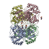
| ||||||||
|---|---|---|---|---|---|---|---|---|---|
| 1 | 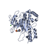
| ||||||||
| 2 | 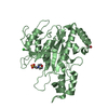
| ||||||||
| 3 | 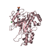
| ||||||||
| 4 | 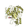
| ||||||||
| Unit cell |
|
-Components
| #1: Protein | Mass: 37952.562 Da / Num. of mol.: 4 / Mutation: D3C,G283C Source method: isolated from a genetically manipulated source Source: (gene. exp.) Streptomyces mobaraensis (bacteria) / Production host: Corynebacterium glutamicum (bacteria)References: UniProt: P81453, protein-glutamine gamma-glutamyltransferase #2: Chemical | ChemComp-MES /   Mass: 195.237 Da / Num. of mol.: 4 / Source method: obtained synthetically / Formula: C6H13NO4S / Comment: pH buffer*YM Mass: 195.237 Da / Num. of mol.: 4 / Source method: obtained synthetically / Formula: C6H13NO4S / Comment: pH buffer*YM#3: Chemical | ChemComp-CL /   Mass: 35.453 Da / Num. of mol.: 4 / Source method: obtained synthetically / Formula: Cl Mass: 35.453 Da / Num. of mol.: 4 / Source method: obtained synthetically / Formula: Cl#4: Chemical | ChemComp-PEG / |   Mass: 106.120 Da / Num. of mol.: 1 / Source method: obtained synthetically / Formula: C4H10O3 Mass: 106.120 Da / Num. of mol.: 1 / Source method: obtained synthetically / Formula: C4H10O3#5: Water | ChemComp-HOH / |  Mass: 18.015 Da / Num. of mol.: 1106 / Source method: isolated from a natural source / Formula: H2O Mass: 18.015 Da / Num. of mol.: 1106 / Source method: isolated from a natural source / Formula: H2OHas ligand of interest | N | Has protein modification | Y | |
|---|
-Experimental details
-Experiment
| Experiment | Method: X-RAY DIFFRACTION / Number of used crystals: 1 |
|---|
- Sample preparation
| Crystal | Density Matthews: 2.39 Å3/Da / Density % sol: 48.64 % |
|---|---|
| Crystal grow | Temperature: 293 K / Method: vapor diffusion, hanging drop / pH: 5 Details: 25 w/v% polyethylene glycol 1000, 100 mM MES-NaOH buffer (pH5.0), and 25 mM CaCl2 |
-Data collection
| Diffraction | Mean temperature: 100 K / Serial crystal experiment: N |
|---|---|
| Diffraction source | Source: SYNCHROTRON / Site: Photon Factory / Beamline: AR-NW12A / Wavelength: 1 Å |
| Detector | Type: ADSC QUANTUM 210 / Detector: CCD / Date: Feb 18, 2010 |
| Radiation | Protocol: SINGLE WAVELENGTH / Monochromatic (M) / Laue (L): M / Scattering type: x-ray |
| Radiation wavelength | Wavelength: 1 Å / Relative weight: 1 |
| Reflection | Resolution: 2→78.659 Å / Num. obs: 93774 / % possible obs: 97.1 % / Redundancy: 3.8288 % / Rmerge(I) obs: 0.037 / Net I/σ(I): 24.04 |
| Reflection shell | Resolution: 2→2.2 Å / Rmerge(I) obs: 0.143 / Num. unique obs: 22950 |
- Processing
| Software |
| |||||||||||||||||||||||||||||||||||||||||||||||||||||||||||||||||||||||||||||||||||||||||||||||||||||||||||||||||||||||||||||||||||||||||||||||||||
|---|---|---|---|---|---|---|---|---|---|---|---|---|---|---|---|---|---|---|---|---|---|---|---|---|---|---|---|---|---|---|---|---|---|---|---|---|---|---|---|---|---|---|---|---|---|---|---|---|---|---|---|---|---|---|---|---|---|---|---|---|---|---|---|---|---|---|---|---|---|---|---|---|---|---|---|---|---|---|---|---|---|---|---|---|---|---|---|---|---|---|---|---|---|---|---|---|---|---|---|---|---|---|---|---|---|---|---|---|---|---|---|---|---|---|---|---|---|---|---|---|---|---|---|---|---|---|---|---|---|---|---|---|---|---|---|---|---|---|---|---|---|---|---|---|---|---|---|---|
| Refinement | Method to determine structure: MOLECULAR REPLACEMENT / Resolution: 2→47.08 Å / Cor.coef. Fo:Fc: 0.953 / Cor.coef. Fo:Fc free: 0.924 / SU B: 4.3 / SU ML: 0.121 / Cross valid method: FREE R-VALUE / ESU R: 0.199 / ESU R Free: 0.176 / Details: Hydrogens have not been used
| |||||||||||||||||||||||||||||||||||||||||||||||||||||||||||||||||||||||||||||||||||||||||||||||||||||||||||||||||||||||||||||||||||||||||||||||||||
| Solvent computation | Ion probe radii: 0.8 Å / Shrinkage radii: 0.8 Å / VDW probe radii: 1.2 Å / Solvent model: MASK BULK SOLVENT | |||||||||||||||||||||||||||||||||||||||||||||||||||||||||||||||||||||||||||||||||||||||||||||||||||||||||||||||||||||||||||||||||||||||||||||||||||
| Displacement parameters | Biso mean: 30.099 Å2
| |||||||||||||||||||||||||||||||||||||||||||||||||||||||||||||||||||||||||||||||||||||||||||||||||||||||||||||||||||||||||||||||||||||||||||||||||||
| Refinement step | Cycle: LAST / Resolution: 2→47.08 Å
| |||||||||||||||||||||||||||||||||||||||||||||||||||||||||||||||||||||||||||||||||||||||||||||||||||||||||||||||||||||||||||||||||||||||||||||||||||
| Refine LS restraints |
| |||||||||||||||||||||||||||||||||||||||||||||||||||||||||||||||||||||||||||||||||||||||||||||||||||||||||||||||||||||||||||||||||||||||||||||||||||
| LS refinement shell |
|
