Resolution: 2.8→25.765 Å / Cor.coef. Fo:Fc: 0.834 / Cor.coef. Fo:Fc free: 0.724 / Cross valid method: FREE R-VALUE Details: Hydrogens have been used if present in the input file
Rfactor
Num. reflection
% reflection
Rfree
0.3261
129
5.025 %
Rwork
0.2943
2438
-
all
0.296
-
-
obs
-
2567
80.698 %
Solvent computation
Ion probe radii: 0.8 Å / Shrinkage radii: 0.8 Å / VDW probe radii: 1.2 Å / Solvent model: MASK BULK SOLVENT
Displacement parameters
Biso mean: 35.48 Å2
Baniso -1
Baniso -2
Baniso -3
1-
-4.552 Å2
0 Å2
0 Å2
2-
-
-4.552 Å2
0 Å2
3-
-
-
9.103 Å2
Refine LS restraints
Refine-ID
Type
Dev ideal
Dev ideal target
Number
ELECTRONCRYSTALLOGRAPHY
r_bond_refined_d
0.004
0.012
1021
ELECTRONCRYSTALLOGRAPHY
r_angle_refined_deg
1.315
1.777
1381
ELECTRONCRYSTALLOGRAPHY
r_dihedral_angle_1_deg
6.239
5
128
ELECTRONCRYSTALLOGRAPHY
r_dihedral_angle_2_deg
3.047
5
11
ELECTRONCRYSTALLOGRAPHY
r_dihedral_angle_3_deg
14.827
10
166
ELECTRONCRYSTALLOGRAPHY
r_dihedral_angle_6_deg
14.487
10
50
ELECTRONCRYSTALLOGRAPHY
r_chiral_restr
0.098
0.2
144
ELECTRONCRYSTALLOGRAPHY
r_gen_planes_refined
0.005
0.02
819
ELECTRONCRYSTALLOGRAPHY
r_nbd_refined
0.253
0.2
509
ELECTRONCRYSTALLOGRAPHY
r_nbtor_refined
0.31
0.2
698
ELECTRONCRYSTALLOGRAPHY
r_xyhbond_nbd_refined
0.45
0.2
50
ELECTRONCRYSTALLOGRAPHY
r_symmetry_nbd_refined
0.2
0.2
24
ELECTRONCRYSTALLOGRAPHY
r_symmetry_xyhbond_nbd_refined
0.279
0.2
11
ELECTRONCRYSTALLOGRAPHY
r_mcbond_it
2.485
3.352
515
ELECTRONCRYSTALLOGRAPHY
r_mcangle_it
4.164
6.02
642
ELECTRONCRYSTALLOGRAPHY
r_scbond_it
3.029
3.772
506
ELECTRONCRYSTALLOGRAPHY
r_scangle_it
4.984
6.786
739
ELECTRONCRYSTALLOGRAPHY
r_lrange_it
9.83
41.305
4456
LS refinement shell
Refine-ID: ELECTRON CRYSTALLOGRAPHY / Total num. of bins used: 20
Resolution (Å)
Rfactor Rfree
Num. reflection Rfree
Rfactor Rwork
Num. reflection Rwork
Rfactor all
Num. reflection all
Fsc free
Fsc work
% reflection obs (%)
WRfactor Rwork
2.801-2.873
0.358
9
0.362
161
0.362
215
0.891
0.865
79.0698
0.332
2.873-2.95
0.186
5
0.338
173
0.332
223
0.991
0.871
79.8206
0.294
2.95-3.035
0.196
7
0.296
170
0.291
211
0.952
0.902
83.8863
0.261
3.035-3.127
0.467
8
0.306
166
0.311
212
0.781
0.901
82.0755
0.264
3.127-3.228
0.235
9
0.264
147
0.262
200
0.96
0.919
78
0.22
3.228-3.339
0.229
10
0.25
149
0.249
192
0.953
0.925
82.8125
0.204
3.339-3.463
0.397
10
0.244
154
0.253
201
0.925
0.935
81.592
0.203
3.463-3.602
0.24
7
0.223
136
0.224
177
0.98
0.951
80.791
0.18
3.602-3.758
0.372
11
0.262
136
0.271
182
0.858
0.935
80.7692
0.217
3.758-3.938
0.358
4
0.219
131
0.222
165
0.991
0.945
81.8182
0.166
3.938-4.145
0.542
1
0.245
127
0.247
161
0.935
79.5031
0.178
4.145-4.39
0.275
7
0.23
125
0.233
157
0.922
0.949
84.0764
0.185
4.39-4.683
0.355
7
0.264
106
0.271
146
0.916
0.945
77.3973
0.218
4.683-5.044
0.174
5
0.281
111
0.275
139
0.996
0.926
83.4532
0.199
5.044-5.505
0.194
3
0.382
102
0.373
136
0.995
0.878
77.2059
0.276
5.505-6.12
0.402
4
0.328
95
0.331
116
0.921
0.893
85.3448
0.248
6.12-7.001
0.598
8
0.394
76
0.413
106
0.843
0.877
79.2453
0.298
7.001-8.42
0.334
6
0.342
72
0.341
96
0.904
0.889
81.25
0.274
8.42-11.319
0.337
4
0.475
61
0.466
82
0.862
0.817
79.2683
0.359
11.319-25.765
0.502
4
0.569
40
0.565
57
0.35
0.827
77.193
0.445
+
About Yorodumi
-
News
-
Feb 9, 2022. New format data for meta-information of EMDB entries
New format data for meta-information of EMDB entries
Version 3 of the EMDB header file is now the official format.
The previous official version 1.9 will be removed from the archive.
In the structure databanks used in Yorodumi, some data are registered as the other names, "COVID-19 virus" and "2019-nCoV". Here are the details of the virus and the list of structure data.
Jan 31, 2019. EMDB accession codes are about to change! (news from PDBe EMDB page)
EMDB accession codes are about to change! (news from PDBe EMDB page)
The allocation of 4 digits for EMDB accession codes will soon come to an end. Whilst these codes will remain in use, new EMDB accession codes will include an additional digit and will expand incrementally as the available range of codes is exhausted. The current 4-digit format prefixed with “EMD-” (i.e. EMD-XXXX) will advance to a 5-digit format (i.e. EMD-XXXXX), and so on. It is currently estimated that the 4-digit codes will be depleted around Spring 2019, at which point the 5-digit format will come into force.
The EM Navigator/Yorodumi systems omit the EMD- prefix.
Related info.:Q: What is EMD? / ID/Accession-code notation in Yorodumi/EM Navigator
Yorodumi is a browser for structure data from EMDB, PDB, SASBDB, etc.
This page is also the successor to EM Navigator detail page, and also detail information page/front-end page for Omokage search.
The word "yorodu" (or yorozu) is an old Japanese word meaning "ten thousand". "mi" (miru) is to see.
Related info.:EMDB / PDB / SASBDB / Comparison of 3 databanks / Yorodumi Search / Aug 31, 2016. New EM Navigator & Yorodumi / Yorodumi Papers / Jmol/JSmol / Function and homology information / Changes in new EM Navigator and Yorodumi
 Movie
Movie Controller

 Yorodumi
 Open data
 Basic information
 Components
 Keywords
 Function and homology information
 MOLECULAR REPLACEMENT / cryo EM / Resolution: 2.8 Å
  Authors
 Citation
 Structure visualization
 Downloads & links
 Other downloads
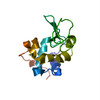
 PDBj
PDBj


 Assembly
 Mass: 18.015 Da / Num. of mol.: 12 / Source method: isolated from a natural source / Formula: H2O
 Sample preparation
 Processing