 Movie
Movie Controller

[English] 日本語
 Yorodumi
- PDB-9iab: Structure of the Argonaute-associated Cas4 family protein 1 (ACE1... -
+ Open data

- Basic information
| Entry | Database: PDB / ID: 9iab | ||||||||||||||||||
|---|---|---|---|---|---|---|---|---|---|---|---|---|---|---|---|---|---|---|---|
| Title | Structure of the Argonaute-associated Cas4 family protein 1 (ACE1) from Chroococcidiopsis thermalis (CtACE1) | ||||||||||||||||||
 Components Components | PD-(D/E)XK endonuclease-like domain-containing protein | ||||||||||||||||||
 Keywords Keywords | IMMUNE SYSTEM / Cas4 family protein / deoxyribonuclease / hydrolase | ||||||||||||||||||
| Function / homology | PD-(D/E)XK endonuclease-like domain, AddAB-type / PD-(D/E)XK nuclease superfamily / PD-(D/E)XK endonuclease-like domain superfamily / IRON/SULFUR CLUSTER / PD-(D/E)XK endonuclease-like domain-containing protein Function and homology information Function and homology information | ||||||||||||||||||
| Biological species |  Chroococcidiopsis thermalis (bacteria) Chroococcidiopsis thermalis (bacteria) | ||||||||||||||||||
| Method |  X-RAY DIFFRACTION / SYNCHROTRON / SAD / Resolution: 1.57 Å X-RAY DIFFRACTION / SYNCHROTRON / SAD / Resolution: 1.57 Å | ||||||||||||||||||
 Authors Authors | Bobadilla Ugarte, P. / Halter, S. / Jinek, M. / Swarts, D.C. | ||||||||||||||||||
| Funding support | European Union,  Netherlands, Netherlands,  Mexico, 5items Mexico, 5items
| ||||||||||||||||||
 Citation Citation | Journal: Mol.Cell / Year: 2025 Title: Cyanobacterial Argonautes and Cas4 family nucleases cooperate to interfere with invading DNA. Authors: Bobadilla Ugarte, P. / Halter, S. / Mutte, S.K. / Heijstek, C. / Niault, T. / Terenin, I. / Barendse, P. / Koopal, B. / Roosjen, M. / Boeren, S. / Hauryliuk, V. / Jinek, M. / Westphal, A.H. / Swarts, D.C. | ||||||||||||||||||
| History |
|
- Structure visualization
| Structure viewer | Molecule: MolmilJmol/JSmol |
|---|
- Downloads & links
-Download
| PDBx/mmCIF format | 9iab.cif.gz | 347.7 KB | Display | PDBx/mmCIF format |
|---|---|---|---|---|
| PDB format | pdb9iab.ent.gz | 277.6 KB | Display | PDB format |
| PDBx/mmJSON format | 9iab.json.gz | Tree view | PDBx/mmJSON format | |
| Others |  Other downloads Other downloads |
-Validation report
| Arichive directory | https://data.pdbj.org/pub/pdb/validation_reports/ia/9iabftp://data.pdbj.org/pub/pdb/validation_reports/ia/9iab | HTTPS FTP |
|---|
-Related structure data


-Links
 PDBj
PDBj
- Assembly
| Deposited unit | 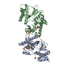
| ||||||||||||
|---|---|---|---|---|---|---|---|---|---|---|---|---|---|
| 1 | 
| ||||||||||||
| 2 | 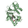
| ||||||||||||
| Unit cell |
|
-Components
| #1: Protein | Mass: 31406.740 Da / Num. of mol.: 2 Source method: isolated from a genetically manipulated source Details: Expression constructed contained His-MBP-TEV site, hence altered amino acids on positions 1-2. Source: (gene. exp.) Chroococcidiopsis thermalis (bacteria) / Gene: Chro_5200 / Production host: #2: Chemical |   Mass: 351.640 Da / Num. of mol.: 2 / Source method: obtained synthetically / Formula: Fe4S4 Mass: 351.640 Da / Num. of mol.: 2 / Source method: obtained synthetically / Formula: Fe4S4#3: Chemical | ChemComp-MES /   Mass: 195.237 Da / Num. of mol.: 4 / Source method: obtained synthetically / Formula: C6H13NO4S / Comment: pH buffer*YM Mass: 195.237 Da / Num. of mol.: 4 / Source method: obtained synthetically / Formula: C6H13NO4S / Comment: pH buffer*YM#4: Chemical | ChemComp-SO4 /   Mass: 96.063 Da / Num. of mol.: 5 / Source method: obtained synthetically / Formula: SO4 Mass: 96.063 Da / Num. of mol.: 5 / Source method: obtained synthetically / Formula: SO4#5: Water | ChemComp-HOH / |  Mass: 18.015 Da / Num. of mol.: 395 / Source method: isolated from a natural source / Formula: H2O Mass: 18.015 Da / Num. of mol.: 395 / Source method: isolated from a natural source / Formula: H2OHas ligand of interest | N | Has protein modification | N | |
|---|
-Experimental details
-Experiment
| Experiment | Method: X-RAY DIFFRACTION / Number of used crystals: 1 |
|---|
- Sample preparation
| Crystal | Density Matthews: 2.24 Å3/Da / Density % sol: 45.19 % |
|---|---|
| Crystal grow | Temperature: 293 K / Method: vapor diffusion, hanging drop Details: Protein solution: -7.5 mg/ml CtACE1 -10 mM HEPES-KOH pH7.5 -250 mM KCl -1 mM DTT Reservoir solution: -0.1 M MES pH 6-7 -0.2 M (NH4)2SO4 -20-25% PEG MME 5K PH range: 6-7 |
-Data collection
| Diffraction | Mean temperature: 100 K / Serial crystal experiment: N |
|---|---|
| Diffraction source | Source: SYNCHROTRON / Site: SLS  / Beamline: X06DA / Wavelength: 1.00003 Å / Beamline: X06DA / Wavelength: 1.00003 Å |
| Detector | Type: DECTRIS PILATUS 2M-F / Detector: PIXEL / Date: May 21, 2017 |
| Radiation | Protocol: SINGLE WAVELENGTH / Monochromatic (M) / Laue (L): M / Scattering type: x-ray |
| Radiation wavelength | Wavelength: 1.00003 Å / Relative weight: 1 |
| Reflection | Resolution: 1.57→43.02 Å / Num. obs: 144407 / % possible obs: 99.29 % / Redundancy: 7 % / CC1/2: 0.999 / Rrim(I) all: 0.034 / Net I/σ(I): 31.65 |
| Reflection shell | Resolution: 1.57→1.63 Å / Redundancy: 7.1 % / Num. unique obs: 7241 / CC1/2: 0.918 / Rrim(I) all: 0.559 / % possible all: 94.57 |
- Processing
| Software |
| |||||||||||||||||||||||||||||||||||||||||||||||||||||||||||||||||||||||||||||||||||||||||||||||||||||||||||||||||||||||||||||||||||||||||||||||||||||||||||||||||||||||||||||||||||||||||||||||||||||||||||
|---|---|---|---|---|---|---|---|---|---|---|---|---|---|---|---|---|---|---|---|---|---|---|---|---|---|---|---|---|---|---|---|---|---|---|---|---|---|---|---|---|---|---|---|---|---|---|---|---|---|---|---|---|---|---|---|---|---|---|---|---|---|---|---|---|---|---|---|---|---|---|---|---|---|---|---|---|---|---|---|---|---|---|---|---|---|---|---|---|---|---|---|---|---|---|---|---|---|---|---|---|---|---|---|---|---|---|---|---|---|---|---|---|---|---|---|---|---|---|---|---|---|---|---|---|---|---|---|---|---|---|---|---|---|---|---|---|---|---|---|---|---|---|---|---|---|---|---|---|---|---|---|---|---|---|---|---|---|---|---|---|---|---|---|---|---|---|---|---|---|---|---|---|---|---|---|---|---|---|---|---|---|---|---|---|---|---|---|---|---|---|---|---|---|---|---|---|---|---|---|---|---|---|---|---|
| Refinement | Method to determine structure: SAD / Resolution: 1.57→43.02 Å / SU ML: 0.1826 / Cross valid method: FREE R-VALUE / σ(F): 1.37 / Phase error: 22.815 Stereochemistry target values: GeoStd + Monomer Library + CDL v1.2
| |||||||||||||||||||||||||||||||||||||||||||||||||||||||||||||||||||||||||||||||||||||||||||||||||||||||||||||||||||||||||||||||||||||||||||||||||||||||||||||||||||||||||||||||||||||||||||||||||||||||||||
| Solvent computation | Shrinkage radii: 0.9 Å / VDW probe radii: 1.1 Å / Solvent model: FLAT BULK SOLVENT MODEL | |||||||||||||||||||||||||||||||||||||||||||||||||||||||||||||||||||||||||||||||||||||||||||||||||||||||||||||||||||||||||||||||||||||||||||||||||||||||||||||||||||||||||||||||||||||||||||||||||||||||||||
| Displacement parameters | Biso mean: 36.4 Å2 | |||||||||||||||||||||||||||||||||||||||||||||||||||||||||||||||||||||||||||||||||||||||||||||||||||||||||||||||||||||||||||||||||||||||||||||||||||||||||||||||||||||||||||||||||||||||||||||||||||||||||||
| Refinement step | Cycle: LAST / Resolution: 1.57→43.02 Å
| |||||||||||||||||||||||||||||||||||||||||||||||||||||||||||||||||||||||||||||||||||||||||||||||||||||||||||||||||||||||||||||||||||||||||||||||||||||||||||||||||||||||||||||||||||||||||||||||||||||||||||
| Refine LS restraints |
| |||||||||||||||||||||||||||||||||||||||||||||||||||||||||||||||||||||||||||||||||||||||||||||||||||||||||||||||||||||||||||||||||||||||||||||||||||||||||||||||||||||||||||||||||||||||||||||||||||||||||||
| LS refinement shell |
|
