 Movie
Movie Controller

[English] 日本語
 Yorodumi
- PDB-9i55: Mouse phosphomannomutase 2 in complex with the activator glucose-... -
+ Open data

- Basic information
| Entry | Database: PDB / ID: 9i55 | |||||||||
|---|---|---|---|---|---|---|---|---|---|---|
| Title | Mouse phosphomannomutase 2 in complex with the activator glucose-1,6-bisphosphate | |||||||||
 Components Components | Phosphomannomutase 2 | |||||||||
 Keywords Keywords | ISOMERASE / N-glycosylation / congenital disorder of glycosylation / carbohydrate-deficient / hypoglycosylation / homodimer / phosphoglucomutase / nucleotide-sugar biosynthesis / mannose-1-phosphate / GDP-mannose | |||||||||
| Function / homology |  Function and homology information Function and homology informationSynthesis of GDP-mannose / : / : / phosphomannomutase / phosphomannomutase activity / GDP-mannose biosynthetic process / mannose metabolic process / protein N-linked glycosylation / microtubule cytoskeleton / cilium ...Synthesis of GDP-mannose / : / : / phosphomannomutase / phosphomannomutase activity / GDP-mannose biosynthetic process / mannose metabolic process / protein N-linked glycosylation / microtubule cytoskeleton / cilium / neuronal cell body / nucleoplasm / metal ion binding / cytoplasm / cytosol Similarity search - Function | |||||||||
| Biological species |  | |||||||||
| Method |  X-RAY DIFFRACTION / SYNCHROTRON / MOLECULAR REPLACEMENT / Resolution: 2.75 Å X-RAY DIFFRACTION / SYNCHROTRON / MOLECULAR REPLACEMENT / Resolution: 2.75 Å | |||||||||
 Authors Authors | Del Cano-Ochoa, F. / Vilar, M. / Vilas, A. / Company, R. / Perez, B. / Ramon-Maiques, S. | |||||||||
| Funding support |  Spain, 2items Spain, 2items
| |||||||||
 Citation Citation | Journal: To Be Published Title: High conformational flexibility of phosphomannomutase 2: Implications for functioning mechanisms, stability and pharmacological chaperone design Authors: Del Cano-Ochoa, F. / Vilar, M. / Vilas, A. / Company, R. / Perez, B. / Ramon-Maiques, S. | |||||||||
| History |
|
- Structure visualization
| Structure viewer | Molecule: MolmilJmol/JSmol |
|---|
- Downloads & links
-Download
| PDBx/mmCIF format | 9i55.cif.gz | 672.3 KB | Display | PDBx/mmCIF format |
|---|---|---|---|---|
| PDB format | pdb9i55.ent.gz | 451.4 KB | Display | PDB format |
| PDBx/mmJSON format | 9i55.json.gz | Tree view | PDBx/mmJSON format | |
| Others |  Other downloads Other downloads |
-Validation report
| Arichive directory | https://data.pdbj.org/pub/pdb/validation_reports/i5/9i55ftp://data.pdbj.org/pub/pdb/validation_reports/i5/9i55 | HTTPS FTP |
|---|
-Related structure data


-Links
 PDBj
PDBj- Assembly
| Deposited unit | 
| ||||||||||||
|---|---|---|---|---|---|---|---|---|---|---|---|---|---|
| 1 | 
| ||||||||||||
| 2 | 
| ||||||||||||
| 3 | 
| ||||||||||||
| Unit cell |
|
-Components
-Protein / Sugars , 2 types, 10 molecules ABDFCE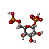
| #1: Protein | Mass: 27848.814 Da / Num. of mol.: 6 Source method: isolated from a genetically manipulated source Details: First two amino acids, "GP", remain from the fusion tag after protease cleavage. Source: (gene. exp.)  #6: Sugar | ChemComp-G16 /  Type: D-saccharide / Mass: 339.108 Da / Num. of mol.: 4 / Source method: obtained synthetically / Formula: C6H13O12P2 / Feature type: SUBJECT OF INVESTIGATION Type: D-saccharide / Mass: 339.108 Da / Num. of mol.: 4 / Source method: obtained synthetically / Formula: C6H13O12P2 / Feature type: SUBJECT OF INVESTIGATION |
|---|
-Non-polymers , 5 types, 312 molecules 


| #2: Chemical | ChemComp-GOL /  Mass: 92.094 Da / Num. of mol.: 1 / Source method: obtained synthetically / Formula: C3H8O3 Mass: 92.094 Da / Num. of mol.: 1 / Source method: obtained synthetically / Formula: C3H8O3 | ||||||
|---|---|---|---|---|---|---|---|
| #3: Chemical | ChemComp-MG /  Mass: 24.305 Da / Num. of mol.: 12 / Source method: obtained synthetically / Formula: Mg Mass: 24.305 Da / Num. of mol.: 12 / Source method: obtained synthetically / Formula: Mg#4: Chemical | ChemComp-CL /  Mass: 35.453 Da / Num. of mol.: 4 / Source method: obtained synthetically / Formula: Cl Mass: 35.453 Da / Num. of mol.: 4 / Source method: obtained synthetically / Formula: Cl#5: Chemical | ChemComp-NA / |  Mass: 22.990 Da / Num. of mol.: 1 / Source method: obtained synthetically / Formula: Na Mass: 22.990 Da / Num. of mol.: 1 / Source method: obtained synthetically / Formula: Na#7: Water | ChemComp-HOH / | Mass: 18.015 Da / Num. of mol.: 294 / Source method: isolated from a natural source / Formula: H2O |
-Details
| Has ligand of interest | Y |
|---|---|
| Has protein modification | N |
-Experimental details
-Experiment
| Experiment | Method: X-RAY DIFFRACTION / Number of used crystals: 1 |
|---|
- Sample preparation
| Crystal | Density Matthews: 2.58 Å3/Da / Density % sol: 52.39 % |
|---|---|
| Crystal grow | Temperature: 291 K / Method: vapor diffusion Details: Drops of 2 ul of protein and 2 mM glucose-1,6-bisphosphate (Sigma) and 2 ul of reservoir solution. Best crystals appeared after 2-7 days in 0.1 M Tris-HCl pH 8.5, 0.2 M MgCl2, 20-22% PEG ...Details: Drops of 2 ul of protein and 2 mM glucose-1,6-bisphosphate (Sigma) and 2 ul of reservoir solution. Best crystals appeared after 2-7 days in 0.1 M Tris-HCl pH 8.5, 0.2 M MgCl2, 20-22% PEG 8000. Crystals were cryo-protected by briefly soaking in mother liquor with increasing concentrations of PEG up to 30% and 5% glycerol. |
-Data collection
| Diffraction | Mean temperature: 100 K / Serial crystal experiment: N |
|---|---|
| Diffraction source | Source: SYNCHROTRON / Site: ALBA / Beamline: XALOC / Wavelength: 0.97926 Å |
| Detector | Type: DECTRIS PILATUS 6M / Detector: PIXEL / Date: Apr 22, 2022 |
| Radiation | Protocol: SINGLE WAVELENGTH / Monochromatic (M) / Laue (L): M / Scattering type: x-ray |
| Radiation wavelength | Wavelength: 0.97926 Å / Relative weight: 1 |
| Reflection | Resolution: 2.75→213.47 Å / Num. obs: 45273 / % possible obs: 98 % / Redundancy: 6.6 % / Biso Wilson estimate: 61.1 Å2 / CC1/2: 0.993 / Rpim(I) all: 0.097 / Net I/σ(I): 6.6 |
| Reflection shell | Resolution: 2.75→2.85 Å / Redundancy: 6.6 % / Mean I/σ(I) obs: 1 / Num. unique obs: 4408 / CC1/2: 0.558 / Rpim(I) all: 0.883 / % possible all: 100 |
- Processing
| Software |
| |||||||||||||||||||||||||||||||||||||||||||||||||||||||||||||||||||||||||||||||||||||||||||||||||||||||||||||||||||||||||||||||||||||||||||||||||||||||||||||||||||||||||||||||||||||||||||||||||||||||||||||||||||||||||
|---|---|---|---|---|---|---|---|---|---|---|---|---|---|---|---|---|---|---|---|---|---|---|---|---|---|---|---|---|---|---|---|---|---|---|---|---|---|---|---|---|---|---|---|---|---|---|---|---|---|---|---|---|---|---|---|---|---|---|---|---|---|---|---|---|---|---|---|---|---|---|---|---|---|---|---|---|---|---|---|---|---|---|---|---|---|---|---|---|---|---|---|---|---|---|---|---|---|---|---|---|---|---|---|---|---|---|---|---|---|---|---|---|---|---|---|---|---|---|---|---|---|---|---|---|---|---|---|---|---|---|---|---|---|---|---|---|---|---|---|---|---|---|---|---|---|---|---|---|---|---|---|---|---|---|---|---|---|---|---|---|---|---|---|---|---|---|---|---|---|---|---|---|---|---|---|---|---|---|---|---|---|---|---|---|---|---|---|---|---|---|---|---|---|---|---|---|---|---|---|---|---|---|---|---|---|---|---|---|---|---|---|---|---|---|---|---|---|---|
| Refinement | Method to determine structure: MOLECULAR REPLACEMENT / Resolution: 2.75→64.67 Å / SU ML: 0.4747 / Cross valid method: FREE R-VALUE / σ(F): 1.9 / Phase error: 30.2492 Stereochemistry target values: GeoStd + Monomer Library + CDL v1.2
| |||||||||||||||||||||||||||||||||||||||||||||||||||||||||||||||||||||||||||||||||||||||||||||||||||||||||||||||||||||||||||||||||||||||||||||||||||||||||||||||||||||||||||||||||||||||||||||||||||||||||||||||||||||||||
| Solvent computation | Shrinkage radii: 0.9 Å / VDW probe radii: 1.1 Å / Solvent model: FLAT BULK SOLVENT MODEL | |||||||||||||||||||||||||||||||||||||||||||||||||||||||||||||||||||||||||||||||||||||||||||||||||||||||||||||||||||||||||||||||||||||||||||||||||||||||||||||||||||||||||||||||||||||||||||||||||||||||||||||||||||||||||
| Displacement parameters | Biso mean: 65.48 Å2 | |||||||||||||||||||||||||||||||||||||||||||||||||||||||||||||||||||||||||||||||||||||||||||||||||||||||||||||||||||||||||||||||||||||||||||||||||||||||||||||||||||||||||||||||||||||||||||||||||||||||||||||||||||||||||
| Refinement step | Cycle: LAST / Resolution: 2.75→64.67 Å
| |||||||||||||||||||||||||||||||||||||||||||||||||||||||||||||||||||||||||||||||||||||||||||||||||||||||||||||||||||||||||||||||||||||||||||||||||||||||||||||||||||||||||||||||||||||||||||||||||||||||||||||||||||||||||
| Refine LS restraints |
| |||||||||||||||||||||||||||||||||||||||||||||||||||||||||||||||||||||||||||||||||||||||||||||||||||||||||||||||||||||||||||||||||||||||||||||||||||||||||||||||||||||||||||||||||||||||||||||||||||||||||||||||||||||||||
| LS refinement shell |
|
