[English] 日本語
Yorodumi
- PDB-9i0c: E3 ubiquitin ligase CBL-B in complex with inhibitor -

+
Open data


ID or keywords:

Loading...

-
Basic information

Entry
Database: PDB / ID: 9i0c
TitleE3 ubiquitin ligase CBL-B in complex with inhibitor
ComponentsE3 ubiquitin-protein ligase CBL-B
KeywordsLIGASE
Function / homology
Function and homology information


regulation of platelet-derived growth factor receptor-alpha signaling pathway / regulation protein catabolic process at postsynapse / NLS-bearing protein import into nucleus / regulation of postsynaptic neurotransmitter receptor internalization / negative regulation of T cell activation / negative regulation of epidermal growth factor receptor signaling pathway / negative regulation of T cell receptor signaling pathway / protein K63-linked ubiquitination / phosphotyrosine residue binding / receptor tyrosine kinase binding ...regulation of platelet-derived growth factor receptor-alpha signaling pathway / regulation protein catabolic process at postsynapse / NLS-bearing protein import into nucleus / regulation of postsynaptic neurotransmitter receptor internalization / negative regulation of T cell activation / negative regulation of epidermal growth factor receptor signaling pathway / negative regulation of T cell receptor signaling pathway / protein K63-linked ubiquitination / phosphotyrosine residue binding / receptor tyrosine kinase binding / RING-type E3 ubiquitin transferase / ubiquitin protein ligase activity / Antigen processing: Ubiquitination & Proteasome degradation / cell surface receptor signaling pathway / postsynapse / protein stabilization / membrane raft / calcium ion binding / glutamatergic synapse / signal transduction / zinc ion binding / plasma membrane / cytosol
Similarity search - Function
E3 ubiquitin-protein ligase CBL-B, RING finger, HC subclass / Adaptor protein Cbl, N-terminal helical / Adaptor protein Cbl, EF hand-like / Adaptor protein Cbl, SH2-like domain / Adaptor protein Cbl, PTB domain / Adaptor protein Cbl / CBL proto-oncogene N-terminal domain 1 / CBL proto-oncogene N-terminus, EF hand-like domain / CBL proto-oncogene N-terminus, SH2-like domain / Cbl-type phosphotyrosine-binding (Cbl-PTB) domain profile. ...E3 ubiquitin-protein ligase CBL-B, RING finger, HC subclass / Adaptor protein Cbl, N-terminal helical / Adaptor protein Cbl, EF hand-like / Adaptor protein Cbl, SH2-like domain / Adaptor protein Cbl, PTB domain / Adaptor protein Cbl / CBL proto-oncogene N-terminal domain 1 / CBL proto-oncogene N-terminus, EF hand-like domain / CBL proto-oncogene N-terminus, SH2-like domain / Cbl-type phosphotyrosine-binding (Cbl-PTB) domain profile. / Adaptor protein Cbl, N-terminal domain superfamily / Ubiquitin associated domain / Ubiquitin-associated domain / Ubiquitin-associated domain (UBA) profile. / Zinc finger, C3HC4 RING-type / Zinc finger, C3HC4 type (RING finger) / Zinc finger, RING-type, conserved site / Zinc finger RING-type signature. / Ring finger / SH2 domain superfamily / Zinc finger RING-type profile. / Zinc finger, RING-type / EF-hand domain pair / Zinc finger, RING/FYVE/PHD-type
Similarity search - Domain/homology
: / E3 ubiquitin-protein ligase CBL-B
Similarity search - Component
Biological speciesHomo sapiens (human)
MethodX-RAY DIFFRACTION / SYNCHROTRON / MOLECULAR REPLACEMENT / Resolution: 2.123 Å
AuthorsSchimpl, M.
Funding support1items
OrganizationGrant numberCountry
Not funded
CitationJournal: To be published
Title: Inhibiting Ubiquitin ligases: A Case Study on the Discovery, Mechanism, and Optimization of Cbl-b Inhibitors
Authors: Chinn, A.J. / Quinn, T.R. / Boerth, J.A. / Kohara, K. / Bommakanti, G.
History
DepositionJan 14, 2025Deposition site: PDBE / Processing site: PDBE
Revision 1.0Jul 29, 2026Provider: repository / Type: Initial release

-
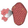
Structure visualization

Structure viewerMolecule:
MolmilJmol/JSmol

Downloads & links

-
Assembly

Deposited unit
A: E3 ubiquitin-protein ligase CBL-B
hetero molecules


Theoretical massNumber of molelcules
Total (without water)46,3525
Polymers45,8101
Non-polymers5424
Water93752
1


  • Idetical with deposited unit
  • defined by author
  • Evidence: gel filtration
TypeNameSymmetry operationNumber
identity operation1_555x,y,z1
Buried area850 Å2
ΔGint3 kcal/mol
Surface area18040 Å2
Unit cell
Length a, b, c (Å)57.183, 74.533, 97.499
Angle α, β, γ (deg.)90.00, 90.00, 90.00
Int Tables number19
Space group name H-MP212121

-
Components

#1: Protein E3 ubiquitin-protein ligase CBL-B


Mass: 45809.512 Da / Num. of mol.: 1
Source method: isolated from a genetically manipulated source
Source: (gene. exp.) Homo sapiens (human) / Gene: CBLB / Production host: Escherichia coli (E. coli) / References: UniProt: Q13191
#2: Chemical ChemComp-A1IZE / 1-[3-[3-methyl-1-(4-methyl-1,2,4-triazol-3-yl)cyclobutyl]phenyl]-5-(trifluoromethyl)pyridin-2-one


Mass: 388.386 Da / Num. of mol.: 1 / Source method: obtained synthetically / Formula: C20H19F3N4O / Feature type: SUBJECT OF INVESTIGATION
#3: Chemical ChemComp-ZN / ZINC ION


Mass: 65.409 Da / Num. of mol.: 2 / Source method: obtained synthetically / Formula: Zn
#4: Chemical ChemComp-NA / SODIUM ION


Mass: 22.990 Da / Num. of mol.: 1 / Source method: obtained synthetically / Formula: Na
#5: Water ChemComp-HOH / water


Mass: 18.015 Da / Num. of mol.: 52 / Source method: isolated from a natural source / Formula: H2O
Has ligand of interestY
Has protein modificationN

-
Experimental details

-
Experiment

ExperimentMethod: X-RAY DIFFRACTION / Number of used crystals: 1

-
Sample preparation

CrystalDensity Matthews: 2.27 Å3/Da / Density % sol: 45.76 %
Crystal growTemperature: 293 K / Method: vapor diffusion, sitting drop / pH: 8
Details: 9 % PEG8000, 0.05 M Mg Acetate, 2.5 % MPD, 0.05 M PCPT pH 8.0

-
Data collection

DiffractionMean temperature: 100 K / Serial crystal experiment: N
Diffraction sourceSource: SYNCHROTRON / Site: SOLEIL / Beamline: PROXIMA 2 / Wavelength: 0.98011 Å
DetectorType: DECTRIS EIGER X 16M / Detector: PIXEL / Date: Nov 11, 2021
RadiationProtocol: SINGLE WAVELENGTH / Monochromatic (M) / Laue (L): M / Scattering type: x-ray
Radiation wavelengthWavelength: 0.98011 Å / Relative weight: 1
ReflectionResolution: 2.12→48.75 Å / Num. obs: 23838 / % possible obs: 97.7 % / Redundancy: 13.3 % / CC1/2: 0.998 / Rmerge(I) obs: 0.122 / Rpim(I) all: 0.035 / Rrim(I) all: 0.127 / Χ2: 0.9 / Net I/σ(I): 13.5 / Num. measured all: 317742
Reflection shellResolution: 2.12→2.17 Å / % possible obs: 74.7 % / Redundancy: 12.3 % / Rmerge(I) obs: 1.517 / Num. measured all: 15922 / Num. unique obs: 1293 / CC1/2: 0.661 / Rpim(I) all: 0.433 / Rrim(I) all: 1.581 / Χ2: 0.59 / Net I/σ(I) obs: 1.3

-
Processing

Software
NameVersionClassification
BUSTER2.11.8 (10-JUL-2024)refinement
Aimlessdata scaling
XDSdata reduction
AMoREphasing
RefinementMethod to determine structure: MOLECULAR REPLACEMENT / Resolution: 2.123→48.75 Å / Cor.coef. Fo:Fc: 0.936 / Cor.coef. Fo:Fc free: 0.921 / SU R Cruickshank DPI: 0.245 / Cross valid method: THROUGHOUT / σ(F): 0 / SU R Blow DPI: 0.244 / SU Rfree Blow DPI: 0.196 / SU Rfree Cruickshank DPI: 0.198
RfactorNum. reflection% reflectionSelection details
Rfree0.2539 1196 5.04 %RANDOM
Rwork0.2137 ---
obs0.2158 23749 98.2 %-
Displacement parametersBiso mean: 53.65 Å2
Baniso -1Baniso -2Baniso -3
1--7.8219 Å20 Å20 Å2
2--10.9741 Å20 Å2
3----3.1522 Å2
Refine analyzeLuzzati coordinate error obs: 0.29 Å
Refinement stepCycle: 1 / Resolution: 2.123→48.75 Å
ProteinNucleic acidLigandSolventTotal
Num. atoms3105 0 31 52 3188
Refine LS restraints
Refine-IDTypeDev idealNumberRestraint functionWeight
X-RAY DIFFRACTIONt_bond_d0.0083211HARMONIC2
X-RAY DIFFRACTIONt_angle_deg0.864347HARMONIC2
X-RAY DIFFRACTIONt_dihedral_angle_d1121SINUSOIDAL2
X-RAY DIFFRACTIONt_incorr_chiral_ct
X-RAY DIFFRACTIONt_pseud_angle
X-RAY DIFFRACTIONt_trig_c_planes
X-RAY DIFFRACTIONt_gen_planes538HARMONIC5
X-RAY DIFFRACTIONt_it3211HARMONIC10
X-RAY DIFFRACTIONt_nbd
X-RAY DIFFRACTIONt_omega_torsion2.83
X-RAY DIFFRACTIONt_other_torsion17.05
X-RAY DIFFRACTIONt_improper_torsion
X-RAY DIFFRACTIONt_chiral_improper_torsion410SEMIHARMONIC5
X-RAY DIFFRACTIONt_sum_occupancies
X-RAY DIFFRACTIONt_utility_distance
X-RAY DIFFRACTIONt_utility_angle
X-RAY DIFFRACTIONt_utility_torsion
X-RAY DIFFRACTIONt_ideal_dist_contact2642SEMIHARMONIC4
LS refinement shellResolution: 2.123→2.15 Å
RfactorNum. reflection% reflection
Rfree0.4114 -4.63 %
Rwork0.3541 453 -
obs--61.45 %
Refinement TLS params.Method: refined / Origin x: 19.8623 Å / Origin y: 15.6316 Å / Origin z: 28.7004 Å
111213212223313233
T-0.0855 Å20.0365 Å20.0265 Å2--0.1223 Å20.0125 Å2---0.0926 Å2
L0.2636 °2-0.1628 °2-0.2468 °2-1.8789 °2-0.0655 °2--1.1165 °2
S0.1221 Å °0.007 Å °-0.037 Å °-0.0209 Å °-0.0514 Å °0.0468 Å °-0.1587 Å °-0.0612 Å °-0.0707 Å °
Refinement TLS groupSelection details: { A|* }

+
About Yorodumi

-
News

-
Feb 9, 2022. New format data for meta-information of EMDB entries

New format data for meta-information of EMDB entries

  • Version 3 of the EMDB header file is now the official format.
  • The previous official version 1.9 will be removed from the archive.

Related info.:EMDB header

External links:wwPDB to switch to version 3 of the EMDB data model

-
Aug 12, 2020. Covid-19 info

Covid-19 info

URL: https://pdbj.org/emnavi/covid19.php

New page: Covid-19 featured information page in EM Navigator.

Related info.:Covid-19 info / Mar 5, 2020. Novel coronavirus structure data

+
Mar 5, 2020. Novel coronavirus structure data

Novel coronavirus structure data

Related info.:Yorodumi Speices / Aug 12, 2020. Covid-19 info

External links:COVID-19 featured content - PDBj / Molecule of the Month (242):Coronavirus Proteases

+
Jan 31, 2019. EMDB accession codes are about to change! (news from PDBe EMDB page)

EMDB accession codes are about to change! (news from PDBe EMDB page)

  • The allocation of 4 digits for EMDB accession codes will soon come to an end. Whilst these codes will remain in use, new EMDB accession codes will include an additional digit and will expand incrementally as the available range of codes is exhausted. The current 4-digit format prefixed with “EMD-” (i.e. EMD-XXXX) will advance to a 5-digit format (i.e. EMD-XXXXX), and so on. It is currently estimated that the 4-digit codes will be depleted around Spring 2019, at which point the 5-digit format will come into force.
  • The EM Navigator/Yorodumi systems omit the EMD- prefix.

Related info.:Q: What is EMD? / ID/Accession-code notation in Yorodumi/EM Navigator

External links:EMDB Accession Codes are Changing Soon! / Contact to PDBj

+
Jul 12, 2017. Major update of PDB

Major update of PDB

  • wwPDB released updated PDB data conforming to the new PDBx/mmCIF dictionary.
  • This is a major update changing the version number from 4 to 5, and with Remediation, in which all the entries are updated.
  • In this update, many items about electron microscopy experimental information are reorganized (e.g. em_software).
  • Now, EM Navigator and Yorodumi are based on the updated data.

External links:wwPDB Remediation / Enriched Model Files Conforming to OneDep Data Standards Now Available in the PDB FTP Archive

-
Yorodumi

Thousand views of thousand structures

  • Yorodumi is a browser for structure data from EMDB, PDB, SASBDB, etc.
  • This page is also the successor to EM Navigator detail page, and also detail information page/front-end page for Omokage search.
  • The word "yorodu" (or yorozu) is an old Japanese word meaning "ten thousand". "mi" (miru) is to see.

Related info.:EMDB / PDB / SASBDB / Comparison of 3 databanks / Yorodumi Search / Aug 31, 2016. New EM Navigator & Yorodumi / Yorodumi Papers / Jmol/JSmol / Function and homology information / Changes in new EM Navigator and Yorodumi

Read more