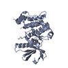
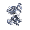
positive regulation of astrocyte activation / positive regulation of microglial cell activation / microtubule associated complex / positive regulation of protein polymerization / tau-protein kinase activity / substantia nigra development / tau protein binding / protein tyrosine kinase activity / learning or memory / non-specific serine/threonine protein kinase ...positive regulation of astrocyte activation / positive regulation of microglial cell activation / microtubule associated complex / positive regulation of protein polymerization / tau-protein kinase activity / substantia nigra development / tau protein binding / protein tyrosine kinase activity / learning or memory / non-specific serine/threonine protein kinase / negative regulation of gene expression / protein serine kinase activity / protein serine/threonine kinase activity / neuronal cell body / positive regulation of gene expression / perinuclear region of cytoplasm / signal transduction / nucleoplasm / ATP binding / nucleus / cytoplasm / cytosol Similarity search - Function
Tau-tubulin kinase 1, catalytic domain / : / Protein kinase domain / Serine/Threonine protein kinases, catalytic domain / Protein kinase, ATP binding site / Protein kinases ATP-binding region signature. / Protein kinase domain profile. / Protein kinase domain / Protein kinase-like domain superfamily Similarity search - Domain/homology
Type: DECTRIS EIGER X 16M / Detector: PIXEL / Date: Feb 10, 2022
Radiation
Protocol: SINGLE WAVELENGTH / Monochromatic (M) / Laue (L): M / Scattering type: x-ray
Radiation wavelength
Wavelength: 0.999995 Å / Relative weight: 1
Reflection
Resolution: 3→48.26 Å / Num. obs: 6759 / % possible obs: 99.8 % / Redundancy: 4.7 % / CC1/2: 0.977 / Net I/σ(I): 6
Reflection shell
Resolution: 3→3.18 Å / Mean I/σ(I) obs: 1.7 / Num. unique obs: 1077 / CC1/2: 0.772
-
Processing
Software
Name
Version
Classification
REFMAC
5.8.0425
refinement
XDS
datareduction
Aimless
datascaling
PHASER
phasing
Refinement
Method to determine structure: MOLECULAR REPLACEMENT / Resolution: 3→48.26 Å / Cor.coef. Fo:Fc: 0.899 / Cor.coef. Fo:Fc free: 0.808 / SU B: 25.236 / SU ML: 0.445 / Cross valid method: THROUGHOUT / ESU R Free: 0.537 / Stereochemistry target values: MAXIMUM LIKELIHOOD / Details: HYDROGENS HAVE BEEN USED IF PRESENT IN THE INPUT
Rfactor
Num. reflection
% reflection
Selection details
Rfree
0.28592
332
4.9 %
RANDOM
Rwork
0.23162
-
-
-
obs
0.23429
6426
99.78 %
-
Solvent computation
Ion probe radii: 0.8 Å / Shrinkage radii: 0.8 Å / VDW probe radii: 1.2 Å / Solvent model: MASK
 Movie
Movie Controller


 Open data
 Basic information
 Components
 Keywords
 Function and homology information
 Homo sapiens (human)
 X-RAY DIFFRACTION /
  Authors
 Canada, 1items
  Citation
 Structure visualization
 Downloads & links
 Other downloads
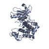


 PDBj
PDBj Assembly

 Mass: 18.015 Da / Num. of mol.: 18 / Source method: isolated from a natural source / Formula: H2O
 Sample preparation
 / Beamline: X06SA / Wavelength: 0.999995 Å
 Processing