 Movie
Movie Controller

[English] 日本語
 Yorodumi
- PDB-9hhb: Crystal Structure of the Plasmodium vivax Bromodomain PvBDP1 in c... -
+ Open data

- Basic information
| Entry | Database: PDB / ID: 9hhb | ||||||
|---|---|---|---|---|---|---|---|
| Title | Crystal Structure of the Plasmodium vivax Bromodomain PvBDP1 in complex with RMM21 | ||||||
 Components Components | Bromo domain-containing protein | ||||||
 Keywords Keywords | TRANSCRIPTION / plasmodium / bromodomain / inhibitor / complex | ||||||
| Function / homology |  Function and homology information Function and homology informationAnkyrin repeats (many copies) / Ankyrin repeat profile. / Ankyrin repeats (3 copies) / Ankyrin repeat region circular profile. / ankyrin repeats / Ankyrin repeat / Ankyrin repeat-containing domain superfamily / Bromodomain, conserved site / Bromodomain signature. / Bromodomain ...Ankyrin repeats (many copies) / Ankyrin repeat profile. / Ankyrin repeats (3 copies) / Ankyrin repeat region circular profile. / ankyrin repeats / Ankyrin repeat / Ankyrin repeat-containing domain superfamily / Bromodomain, conserved site / Bromodomain signature. / Bromodomain / bromo domain / Bromodomain / Bromodomain (BrD) profile. / Bromodomain-like superfamily Similarity search - Domain/homology | ||||||
| Biological species |  | ||||||
| Method |  X-RAY DIFFRACTION / SYNCHROTRON / MOLECULAR REPLACEMENT / Resolution: 2.26 Å X-RAY DIFFRACTION / SYNCHROTRON / MOLECULAR REPLACEMENT / Resolution: 2.26 Å | ||||||
 Authors Authors | Amann, M. / Huegle, M. / Einsle, O. / Guenther, S. | ||||||
| Funding support |  Germany, 1items Germany, 1items
| ||||||
 Citation Citation | Journal: Chemmedchem / Year: 2025 Title: A Novel Inhibitor against the Bromodomain Protein 1 of the Malaria Pathogen Plasmodium Falciparum. Authors: Amann, M. / Warstat, R. / Rechten, K.K. / Theuer, P. / Schustereder, M. / Clavey, S. / Breit, B. / Einsle, O. / Hugle, M. / Petter, M. / Gunther, S. | ||||||
| History |
|
- Structure visualization
| Structure viewer | Molecule: MolmilJmol/JSmol |
|---|
- Downloads & links
-Download
| PDBx/mmCIF format | 9hhb.cif.gz | 68.9 KB | Display | PDBx/mmCIF format |
|---|---|---|---|---|
| PDB format | pdb9hhb.ent.gz | Display | PDB format | |
| PDBx/mmJSON format | 9hhb.json.gz | Tree view | PDBx/mmJSON format | |
| Others |  Other downloads Other downloads |
-Validation report
| Arichive directory | https://data.pdbj.org/pub/pdb/validation_reports/hh/9hhbftp://data.pdbj.org/pub/pdb/validation_reports/hh/9hhb | HTTPS FTP |
|---|
-Related structure data
| Related structure data | 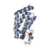 9hgfC 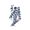 9hh7C  9hh8C  9hhaC  9hhcC  9hhdC C: citing same article ( |
|---|---|
| Similar structure data |
-Links
 PDBj
PDBj- Assembly
| Deposited unit | 
| ||||||||||||
|---|---|---|---|---|---|---|---|---|---|---|---|---|---|
| 1 |
| ||||||||||||
| Unit cell |
| ||||||||||||
| Components on special symmetry positions |
|
-Components
| #1: Protein | Mass: 15163.188 Da / Num. of mol.: 1 Source method: isolated from a genetically manipulated source Source: (gene. exp.) Gene: PVX_111040 / Production host:  |
|---|---|
| #2: Chemical | ChemComp-SO4 /   Mass: 96.063 Da / Num. of mol.: 1 / Source method: obtained synthetically / Formula: SO4 Mass: 96.063 Da / Num. of mol.: 1 / Source method: obtained synthetically / Formula: SO4 |
| #3: Chemical | ChemComp-A1IUY / ~{ Mass: 346.444 Da / Num. of mol.: 1 / Source method: obtained synthetically / Formula: C18H22N2O3S / Feature type: SUBJECT OF INVESTIGATION |
| #4: Water | ChemComp-HOH /  Mass: 18.015 Da / Num. of mol.: 55 / Source method: isolated from a natural source / Formula: H2O Mass: 18.015 Da / Num. of mol.: 55 / Source method: isolated from a natural source / Formula: H2O |
| Has ligand of interest | Y |
| Has protein modification | N |
-Experimental details
-Experiment
| Experiment | Method: X-RAY DIFFRACTION / Number of used crystals: 1 |
|---|
- Sample preparation
| Crystal | Density Matthews: 2.84 Å3/Da / Density % sol: 56.62 % |
|---|---|
| Crystal grow | Temperature: 277 K / Method: vapor diffusion, sitting drop / Details: 35% PEG3350, 0.4 M LiSO4, Tris pH 8,2 |
-Data collection
| Diffraction | Mean temperature: 100 K / Serial crystal experiment: N |
|---|---|
| Diffraction source | Source: SYNCHROTRON / Site: SLS  / Beamline: X06SA / Wavelength: 0.999987 Å / Beamline: X06SA / Wavelength: 0.999987 Å |
| Detector | Type: DECTRIS EIGER X 16M / Detector: PIXEL / Date: Jun 24, 2022 |
| Radiation | Protocol: SINGLE WAVELENGTH / Monochromatic (M) / Laue (L): M / Scattering type: x-ray |
| Radiation wavelength | Wavelength: 0.999987 Å / Relative weight: 1 |
| Reflection | Resolution: 2.26→46.19 Å / Num. obs: 8689 / % possible obs: 100 % / Redundancy: 38.4 % / CC1/2: 0.999 / Rmerge(I) obs: 0.137 / Rpim(I) all: 0.023 / Rrim(I) all: 0.139 / Χ2: 1 / Net I/σ(I): 20 / Num. measured all: 333586 |
| Reflection shell | Resolution: 2.26→2.33 Å / % possible obs: 100 % / Redundancy: 40.8 % / Rmerge(I) obs: 0.965 / Num. measured all: 31733 / Num. unique obs: 778 / CC1/2: 0.975 / Rpim(I) all: 0.153 / Rrim(I) all: 0.977 / Χ2: 1.02 / Net I/σ(I) obs: 5.6 |
- Processing
| Software |
| ||||||||||||||||||||||||||||||||||||||||
|---|---|---|---|---|---|---|---|---|---|---|---|---|---|---|---|---|---|---|---|---|---|---|---|---|---|---|---|---|---|---|---|---|---|---|---|---|---|---|---|---|---|
| Refinement | Method to determine structure: MOLECULAR REPLACEMENT / Resolution: 2.26→46.19 Å / SU ML: 0.35 / Cross valid method: FREE R-VALUE / σ(F): 1.34 / Phase error: 35.55 / Stereochemistry target values: ML
| ||||||||||||||||||||||||||||||||||||||||
| Solvent computation | Shrinkage radii: 0.9 Å / VDW probe radii: 1.1 Å / Solvent model: FLAT BULK SOLVENT MODEL | ||||||||||||||||||||||||||||||||||||||||
| Refinement step | Cycle: LAST / Resolution: 2.26→46.19 Å
| ||||||||||||||||||||||||||||||||||||||||
| Refine LS restraints |
| ||||||||||||||||||||||||||||||||||||||||
| LS refinement shell |
| ||||||||||||||||||||||||||||||||||||||||
| Refinement TLS params. | Method: refined / Origin x: 25.5415 Å / Origin y: -22.8954 Å / Origin z: -0.8998 Å
| ||||||||||||||||||||||||||||||||||||||||
| Refinement TLS group | Selection details: all |
