+ Open data
- Basic information
| Entry | Database: PDB / ID: 9gk1 | |||||||||||||||
|---|---|---|---|---|---|---|---|---|---|---|---|---|---|---|---|---|
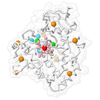
| Title | SSX structure of human cytochrome P450 3A4 at room temperature | |||||||||||||||
 Components Components | Cytochrome P450 3A4 | |||||||||||||||
 Keywords Keywords | OXIDOREDUCTASE / monooxygenase | |||||||||||||||
| Function / homology |  Function and homology information Function and homology informationquinine 3-monooxygenase / 1,8-cineole 2-exo-monooxygenase / albendazole monooxygenase (sulfoxide-forming) / quinine 3-monooxygenase activity / 1,8-cineole 2-exo-monooxygenase activity / 1-alpha,25-dihydroxyvitamin D3 23-hydroxylase activity / vitamin D3 25-hydroxylase activity / vitamin D 24-hydroxylase activity / vitamin D catabolic process / retinoic acid 4-hydroxylase activity ...quinine 3-monooxygenase / 1,8-cineole 2-exo-monooxygenase / albendazole monooxygenase (sulfoxide-forming) / quinine 3-monooxygenase activity / 1,8-cineole 2-exo-monooxygenase activity / 1-alpha,25-dihydroxyvitamin D3 23-hydroxylase activity / vitamin D3 25-hydroxylase activity / vitamin D 24-hydroxylase activity / vitamin D catabolic process / retinoic acid 4-hydroxylase activity / caffeine oxidase activity / estrogen 16-alpha-hydroxylase activity / lipid hydroxylation / aflatoxin metabolic process / anandamide 8,9 epoxidase activity / anandamide 11,12 epoxidase activity / anandamide 14,15 epoxidase activity / testosterone 6-beta-hydroxylase activity / alkaloid catabolic process / Aflatoxin activation and detoxification / Biosynthesis of maresin-like SPMs / monoterpenoid metabolic process / estrogen 2-hydroxylase activity / oxidative demethylation / vitamin D metabolic process / steroid catabolic process / Atorvastatin ADME / steroid hydroxylase activity / Xenobiotics / Phase I - Functionalization of compounds / retinoic acid metabolic process / retinol metabolic process / estrogen metabolic process / unspecific monooxygenase / long-chain fatty acid biosynthetic process / Prednisone ADME / Aspirin ADME / steroid metabolic process / androgen metabolic process / xenobiotic catabolic process / cholesterol metabolic process / steroid binding / xenobiotic metabolic process / monooxygenase activity / oxygen binding / lipid metabolic process / oxidoreductase activity / iron ion binding / intracellular membrane-bounded organelle / heme binding / endoplasmic reticulum membrane / enzyme binding / cytoplasm Similarity search - Function | |||||||||||||||
| Biological species |  Homo sapiens (human) Homo sapiens (human) | |||||||||||||||
| Method |  X-RAY DIFFRACTION / X-RAY DIFFRACTION /  SYNCHROTRON / SYNCHROTRON /  MOLECULAR REPLACEMENT / Resolution: 2.95 Å MOLECULAR REPLACEMENT / Resolution: 2.95 Å | |||||||||||||||
 Authors Authors | Glerup, J. / Branden, G. / Uwangue, O. | |||||||||||||||
| Funding support |  Sweden, 4items Sweden, 4items
| |||||||||||||||
 Citation Citation |  Journal: Arch.Biochem.Biophys. / Year: 2025 Journal: Arch.Biochem.Biophys. / Year: 2025Title: Microcrystallization and room-temperature serial crystallography structure of human cytochrome P450 3A4. Authors: Uwangue, O. / Glerup, J. / Dunge, A. / Bjelcic, M. / Wehlander, G. / Branden, G. | |||||||||||||||
| History |
|
- Structure visualization
| Structure viewer | Molecule:  Molmil Molmil Jmol/JSmol Jmol/JSmol |
|---|
- Downloads & links
- Download
| PDBx/mmCIF format |  9gk1.cif.gz 9gk1.cif.gz | 210.6 KB | Display |  PDBx/mmCIF format PDBx/mmCIF format |
|---|---|---|---|---|
| PDB format |  pdb9gk1.ent.gz pdb9gk1.ent.gz | 168 KB | Display |  PDB format PDB format |
| PDBx/mmJSON format |  9gk1.json.gz 9gk1.json.gz | Tree view |  PDBx/mmJSON format PDBx/mmJSON format | |
| Others |  Other downloads Other downloads |
-Validation report
| Summary document |  9gk1_validation.pdf.gz 9gk1_validation.pdf.gz | 768.6 KB | Display |  wwPDB validaton report wwPDB validaton report |
|---|---|---|---|---|
| Full document |  9gk1_full_validation.pdf.gz 9gk1_full_validation.pdf.gz | 776.4 KB | Display | |
| Data in XML |  9gk1_validation.xml.gz 9gk1_validation.xml.gz | 21.3 KB | Display | |
| Data in CIF |  9gk1_validation.cif.gz 9gk1_validation.cif.gz | 27.5 KB | Display | |
| Arichive directory |  https://data.pdbj.org/pub/pdb/validation_reports/gk/9gk1 https://data.pdbj.org/pub/pdb/validation_reports/gk/9gk1 ftp://data.pdbj.org/pub/pdb/validation_reports/gk/9gk1 ftp://data.pdbj.org/pub/pdb/validation_reports/gk/9gk1 | HTTPS FTP |
-Related structure data
| Similar structure data | Similarity search - Function & homology  F&H Search F&H Search |
|---|
- Links
Links
- Assembly
| Deposited unit | 
| ||||||||
|---|---|---|---|---|---|---|---|---|---|
| 1 |
| ||||||||
| Unit cell |
|
- Components
| #1: Protein | Mass: 55350.289 Da / Num. of mol.: 1 Source method: isolated from a genetically manipulated source Source: (gene. exp.)  Homo sapiens (human) / Gene: CYP3A4, CYP3A3 / Organ: Liver / Production host: Homo sapiens (human) / Gene: CYP3A4, CYP3A3 / Organ: Liver / Production host:  References: UniProt: P08684, unspecific monooxygenase, 1,8-cineole 2-exo-monooxygenase, albendazole monooxygenase (sulfoxide-forming), quinine 3-monooxygenase |
|---|---|
| #2: Chemical | ChemComp-HEM / |
| #3: Water | ChemComp-HOH / |
| Has ligand of interest | Y |
| Has protein modification | N |
-Experimental details
-Experiment
| Experiment | Method:  X-RAY DIFFRACTION / Number of used crystals: 1 X-RAY DIFFRACTION / Number of used crystals: 1 |
|---|
- Sample preparation
| Crystal | Density Matthews: 2.41 Å3/Da / Density % sol: 48.95 % / Description: Square rods |
|---|---|
| Crystal grow | Temperature: 293.15 K / Method: vapor diffusion, sitting drop / pH: 7.3 Details: Final droplet contained 6%v/v PEG3350, 50 mM sodium malonate, 15 mg/mL CYP3A4, 25 mM KPi pH 7.3, 100 mM KCl, 1 mM DTT, 0.5 mM EDTA, 10%v/v glycerol, 1%v/v DMSO and 100 uM beta-napthoflavone. ...Details: Final droplet contained 6%v/v PEG3350, 50 mM sodium malonate, 15 mg/mL CYP3A4, 25 mM KPi pH 7.3, 100 mM KCl, 1 mM DTT, 0.5 mM EDTA, 10%v/v glycerol, 1%v/v DMSO and 100 uM beta-napthoflavone. The droplet was equillibrated against a reservoir of 100 mM sodium malonate and 12%v/v PEG3350. The crystalisation droplet swelled due to high glycerol concentration. |
-Data collection
| Diffraction | Mean temperature: 293 K / Serial crystal experiment: Y |
|---|---|
| Diffraction source | Source:  SYNCHROTRON / Site: SYNCHROTRON / Site:  MAX IV MAX IV  / Beamline: BioMAX / Wavelength: 0.976 Å / Beamline: BioMAX / Wavelength: 0.976 Å |
| Detector | Type: DECTRIS EIGER X 16M / Detector: PIXEL / Date: Dec 9, 2022 Details: BioMAX components: undulator, two front-end beam position monitors, front-end fluorescent screen, optics hutch fixed mask, double-crystal monochromator, fluorescent screen, NanoBPM, slits, ...Details: BioMAX components: undulator, two front-end beam position monitors, front-end fluorescent screen, optics hutch fixed mask, double-crystal monochromator, fluorescent screen, NanoBPM, slits, vertical focusing mirror, horizontal focusing mirror, NanoBPM, beam conditioning unit (BCU), sample and detector. The BCU chamber includes slits, diagnostics, two diamond beam position monitors, attenuators, shutter, slits and diagnostics. |
| Radiation | Monochromator: Si (111) horizontal double crystal monochromator Protocol: SINGLE WAVELENGTH / Monochromatic (M) / Laue (L): M / Scattering type: x-ray |
| Radiation wavelength | Wavelength: 0.976 Å / Relative weight: 1 |
| Reflection | Resolution: 2.95→80.75 Å / Num. obs: 11603 / % possible obs: 100 % / Redundancy: 416.91 % / Biso Wilson estimate: 121.39 Å2 / CC1/2: 0.9953922 / CC star: 0.9988447 / R split: 0.0543 / Net I/σ(I): 14.758752 |
| Reflection shell | Resolution: 2.95→3 Å / Redundancy: 282.9 % / Mean I/σ(I) obs: 1 / Num. unique obs: 547 / CC1/2: 0.5453646 / CC star: 0.8401232 / R split: 0.7304 / % possible all: 100 |
| Serial crystallography measurement | Collection time total: 2 hours / Collimation: Kirkpatrick-Baez mirror pair |
| Serial crystallography sample delivery | Description: Serial X SPINE-mounted Kapton membrane sandwich on a plastic frame Method: fixed target |
| Serial crystallography sample delivery fixed target | Description: 8 x 1 uL crystal slurry on kapton films Details: Arinax Microdiffractomer MD3-down mini-kappagoniometer head Motion control: Arinax Microdiffractomer MD3-down Sample dehydration prevention: Encapsulated by kapton film and glue Sample holding: Serial X SPINE-mounted Kapton membrane sandwich on a plastic frame Support base: SPINE-compatible |
| Serial crystallography data reduction | Frames indexed: 52670 / Frames total: 169142 / Lattices indexed: 66748 |
- Processing
| Software |
| |||||||||||||||||||||||||||||||||||||||||||||||||||||||||||||||||||||||||||||||||||||||||||||||||||||||||||||||||||||||||||||||||||||||||||||||||||||||||||||||||||||||||||||||||||||||||
|---|---|---|---|---|---|---|---|---|---|---|---|---|---|---|---|---|---|---|---|---|---|---|---|---|---|---|---|---|---|---|---|---|---|---|---|---|---|---|---|---|---|---|---|---|---|---|---|---|---|---|---|---|---|---|---|---|---|---|---|---|---|---|---|---|---|---|---|---|---|---|---|---|---|---|---|---|---|---|---|---|---|---|---|---|---|---|---|---|---|---|---|---|---|---|---|---|---|---|---|---|---|---|---|---|---|---|---|---|---|---|---|---|---|---|---|---|---|---|---|---|---|---|---|---|---|---|---|---|---|---|---|---|---|---|---|---|---|---|---|---|---|---|---|---|---|---|---|---|---|---|---|---|---|---|---|---|---|---|---|---|---|---|---|---|---|---|---|---|---|---|---|---|---|---|---|---|---|---|---|---|---|---|---|---|---|---|
| Refinement | Method to determine structure:  MOLECULAR REPLACEMENT / Resolution: 2.95→80.75 Å / Cor.coef. Fo:Fc: 0.946 / Cor.coef. Fo:Fc free: 0.94 / SU B: 65.754 / SU ML: 0.524 / Cross valid method: THROUGHOUT / ESU R Free: 0.519 / Stereochemistry target values: MAXIMUM LIKELIHOOD / Details: HYDROGENS HAVE BEEN ADDED IN THE RIDING POSITIONS MOLECULAR REPLACEMENT / Resolution: 2.95→80.75 Å / Cor.coef. Fo:Fc: 0.946 / Cor.coef. Fo:Fc free: 0.94 / SU B: 65.754 / SU ML: 0.524 / Cross valid method: THROUGHOUT / ESU R Free: 0.519 / Stereochemistry target values: MAXIMUM LIKELIHOOD / Details: HYDROGENS HAVE BEEN ADDED IN THE RIDING POSITIONS
| |||||||||||||||||||||||||||||||||||||||||||||||||||||||||||||||||||||||||||||||||||||||||||||||||||||||||||||||||||||||||||||||||||||||||||||||||||||||||||||||||||||||||||||||||||||||||
| Solvent computation | Ion probe radii: 0.8 Å / Shrinkage radii: 0.8 Å / VDW probe radii: 1.2 Å / Solvent model: MASK | |||||||||||||||||||||||||||||||||||||||||||||||||||||||||||||||||||||||||||||||||||||||||||||||||||||||||||||||||||||||||||||||||||||||||||||||||||||||||||||||||||||||||||||||||||||||||
| Displacement parameters | Biso mean: 143.605 Å2
| |||||||||||||||||||||||||||||||||||||||||||||||||||||||||||||||||||||||||||||||||||||||||||||||||||||||||||||||||||||||||||||||||||||||||||||||||||||||||||||||||||||||||||||||||||||||||
| Refinement step | Cycle: 1 / Resolution: 2.95→80.75 Å
| |||||||||||||||||||||||||||||||||||||||||||||||||||||||||||||||||||||||||||||||||||||||||||||||||||||||||||||||||||||||||||||||||||||||||||||||||||||||||||||||||||||||||||||||||||||||||
| Refine LS restraints |
| |||||||||||||||||||||||||||||||||||||||||||||||||||||||||||||||||||||||||||||||||||||||||||||||||||||||||||||||||||||||||||||||||||||||||||||||||||||||||||||||||||||||||||||||||||||||||
| LS refinement shell | Resolution: 2.95→3.027 Å / Total num. of bins used: 20
| |||||||||||||||||||||||||||||||||||||||||||||||||||||||||||||||||||||||||||||||||||||||||||||||||||||||||||||||||||||||||||||||||||||||||||||||||||||||||||||||||||||||||||||||||||||||||
| Refinement TLS params. | Method: refined / Origin x: -20.331 Å / Origin y: -24.132 Å / Origin z: -13.432 Å
| |||||||||||||||||||||||||||||||||||||||||||||||||||||||||||||||||||||||||||||||||||||||||||||||||||||||||||||||||||||||||||||||||||||||||||||||||||||||||||||||||||||||||||||||||||||||||
| Refinement TLS group |
|
 Movie
Movie Controller


 PDBj
PDBj


