 Movie
Movie Controller


+ Open data

- Basic information
| Entry | Database: PDB / ID: 9gjl | |||||||||||||||||||||||||||
|---|---|---|---|---|---|---|---|---|---|---|---|---|---|---|---|---|---|---|---|---|---|---|---|---|---|---|---|---|
| Title | Entamoeba histolytica Gal/GalNAc lectin bound to LacNAc, mode 2 | |||||||||||||||||||||||||||
 Components Components | (Galactose/N-acetyl-D-galactosamine lectin ...) x 2 | |||||||||||||||||||||||||||
 Keywords Keywords | CELL ADHESION / Entamoeba histolytica / lectin / Gal/GalNAc / trogocytosis | |||||||||||||||||||||||||||
| Function / homology | Galactose-inhibitable lectin 35kDa subunit / Galactose-inhibitable lectin 35 kDa subunit / carbohydrate binding / cell adhesion / plasma membrane / N-acetyl-alpha-lactosamine / Galactose/N-acetyl-D-galactosamine lectin heavy subunit 1 / Galactose/N-acetyl-D-galactosamine lectin light subunit 1 Function and homology information Function and homology information | |||||||||||||||||||||||||||
| Biological species |  Entamoeba histolytica HM-1:IMSS (eukaryote) Entamoeba histolytica HM-1:IMSS (eukaryote) | |||||||||||||||||||||||||||
| Method | ELECTRON MICROSCOPY / single particle reconstruction / cryo EM / Resolution: 3.4 Å | |||||||||||||||||||||||||||
 Authors Authors | Gerard, S.F. / Higgins, M.K. | |||||||||||||||||||||||||||
| Funding support |  United Kingdom, 1items United Kingdom, 1items
| |||||||||||||||||||||||||||
 Citation Citation | Journal: PLoS Pathog / Year: 2026 Title: Structural basis for carbohydrate recognition by the Gal/GalNAc lectin of Entamoeba histolytica involved in host cell adhesion. Authors: Samuel F Gérard / Christina Redfield / Matthew K Higgins / Abstract: Intestinal amoebiasis is caused by Entamoeba histolytica, one of the deadliest human-infective parasites. Central to its pathogenicity is its binding to mucosal carbohydrates, which precedes tissue ...Intestinal amoebiasis is caused by Entamoeba histolytica, one of the deadliest human-infective parasites. Central to its pathogenicity is its binding to mucosal carbohydrates, which precedes tissue damage by trogocytosis. Carbohydrate binding is mediated by a single adhesin, the galactose/N-acetylgalactosamine (Gal/GalNAc) lectin, which is the leading vaccine candidate for amoebiasis. We present the structure of the native heterodimeric lectin, revealing an ordered core containing the light chain and the N-terminal region of the heavy chain. Structures obtained in the presence of ligand show that the Gal/GalNAc binding site is in the light chain, which adopts a β-trefoil fold found in other lectins. An elongated arm emerges from the heavy chain, which adopts multiple positions and may be modulated by sugar binding. This study reveals the molecular basis for sugar binding by the Entamoeba histolytica Gal/GalNAc lectin, a prerequisite for parasite invasion and development of intestinal disease. | |||||||||||||||||||||||||||
| History |
|
- Structure visualization
| Structure viewer | Molecule: MolmilJmol/JSmol |
|---|
- Downloads & links
-Download
| PDBx/mmCIF format | 9gjl.cif.gz | 203 KB | Display | PDBx/mmCIF format |
|---|---|---|---|---|
| PDB format | pdb9gjl.ent.gz | 157.1 KB | Display | PDB format |
| PDBx/mmJSON format | 9gjl.json.gz | Tree view | PDBx/mmJSON format | |
| Others |  Other downloads Other downloads |
-Validation report
| Arichive directory | https://data.pdbj.org/pub/pdb/validation_reports/gj/9gjlftp://data.pdbj.org/pub/pdb/validation_reports/gj/9gjl | HTTPS FTP |
|---|
-Related structure data
| Related structure data |  51396MC  9gedC  9geeC  9gegC  9gehC  9gjaC  9gjbC  9gjcC  9gjhC  9gjiC  9gjjC  9gjkC  9gjmC M: map data used to model this data C: citing same article ( |
|---|---|
| Similar structure data |


-Links
 PDBj
PDBj- Assembly
| Deposited unit | 
|
|---|---|
| 1 |
|
-Components
-Galactose/N-acetyl-D-galactosamine lectin ... , 2 types, 2 molecules HL
| #1: Protein | Mass: 76160.180 Da / Num. of mol.: 1 / Source method: isolated from a natural source Source: (natural) Entamoeba histolytica HM-1:IMSS (eukaryote)References: UniProt: P32022 |
|---|---|
| #2: Protein | Mass: 33637.863 Da / Num. of mol.: 1 / Source method: isolated from a natural source Source: (natural) Entamoeba histolytica HM-1:IMSS (eukaryote)References: UniProt: Q03077 |
-Sugars , 4 types, 5 molecules 
| #3: Polysaccharide | beta-D-mannopyranose-(1-4)-2-acetamido-2-deoxy-beta-D-glucopyranose-(1-4)-2-acetamido-2-deoxy-beta- ...beta-D-mannopyranose-(1-4)-2-acetamido-2-deoxy-beta-D-glucopyranose-(1-4)-2-acetamido-2-deoxy-beta-D-glucopyranose Source method: isolated from a genetically manipulated source |
|---|---|
| #4: Polysaccharide | alpha-D-mannopyranose-(1-2)-alpha-D-mannopyranose-(1-2)-alpha-D-mannopyranose-(1-3)-[alpha-D- ...alpha-D-mannopyranose-(1-2)-alpha-D-mannopyranose-(1-2)-alpha-D-mannopyranose-(1-3)-[alpha-D-mannopyranose-(1-6)]beta-D-mannopyranose-(1-4)-2-acetamido-2-deoxy-beta-D-glucopyranose-(1-4)-2-acetamido-2-deoxy-beta-D-glucopyranose Source method: isolated from a genetically manipulated source |
| #5: Polysaccharide | beta-D-galactopyranose-(1-4)-2-acetamido-2-deoxy-alpha-D-glucopyranose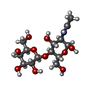  Source method: isolated from a genetically manipulated source Details: oligosaccharide / References: N-acetyl-alpha-lactosamine |
| #6: Sugar |  Type: D-saccharide, beta linking / Mass: 221.208 Da / Num. of mol.: 2 Type: D-saccharide, beta linking / Mass: 221.208 Da / Num. of mol.: 2Source method: isolated from a genetically manipulated source Formula: C8H15NO6 |
-Details
| Has ligand of interest | Y |
|---|---|
| Has protein modification | Y |
-Experimental details
-Experiment
| Experiment | Method: ELECTRON MICROSCOPY |
|---|---|
| EM experiment | Aggregation state: PARTICLE / 3D reconstruction method: single particle reconstruction |
- Sample preparation
| Component | Name: Entamoeba histolytica Gal/GalNAc lectin heavy and light chains Type: COMPLEX / Entity ID: #2 / Source: NATURAL |
|---|---|
| Molecular weight | Experimental value: NO |
| Source (natural) | Organism: Entamoeba histolytica HM-1:IMSS (eukaryote) |
| Buffer solution | pH: 7.5 |
| Specimen | Embedding applied: NO / Shadowing applied: NO / Staining applied: NO / Vitrification applied: YES |
| Vitrification | Cryogen name: ETHANE |
- Electron microscopy imaging
| Experimental equipment |  Model: Titan Krios / Image courtesy: FEI Company |
|---|---|
| Microscopy | Model: FEI TITAN KRIOS |
| Electron gun | Electron source:  FIELD EMISSION GUN / Accelerating voltage: 300 kV / Illumination mode: FLOOD BEAM FIELD EMISSION GUN / Accelerating voltage: 300 kV / Illumination mode: FLOOD BEAM |
| Electron lens | Mode: BRIGHT FIELD / Nominal defocus max: 2500 nm / Nominal defocus min: 1000 nm |
| Image recording | Electron dose: 41.5 e/Å2 / Film or detector model: GATAN K3 (6k x 4k) |
- Processing
| EM software | Name: PHENIX / Category: model refinement | ||||||||||||||||||||||||
|---|---|---|---|---|---|---|---|---|---|---|---|---|---|---|---|---|---|---|---|---|---|---|---|---|---|
| CTF correction | Type: PHASE FLIPPING AND AMPLITUDE CORRECTION | ||||||||||||||||||||||||
| 3D reconstruction | Resolution: 3.4 Å / Resolution method: FSC 0.143 CUT-OFF / Num. of particles: 125174 / Symmetry type: POINT | ||||||||||||||||||||||||
| Refine LS restraints |
|
