[English] 日本語
Yorodumi
- PDB-9gdf: Chloride bound structure of oxidized ba3-type cytochrome c oxidas... -

+
Open data


ID or keywords:

Loading...

-
Basic information

Entry
Database: PDB / ID: 9gdf
TitleChloride bound structure of oxidized ba3-type cytochrome c oxidase confirmed by single-wavelength anomalous diffraction
Components
  • (Cytochrome c oxidase subunit ...) x 2
  • Cytochrome c oxidase polypeptide 2A
KeywordsOXIDOREDUCTASE / Anomalous diffraction / cytochrome c oxidase / high salt / low pH
Function / homology
Function and homology information


cytochrome-c oxidase / oxidative phosphorylation / cytochrome-c oxidase activity / copper ion binding / heme binding / metal ion binding / plasma membrane
Similarity search - Function
Cytochrome c oxidase polypeptide 2A / Cytochrome c oxidase polypeptide 2A, ba3 type / Cytochrome c oxidase subunit IIa family / Ba3-like heme-copper oxidase subunit I / Cytochrome C oxidase subunit IIa, transmembrane domain / Cytochrome C oxidase subunit II, transmembrane / Ba3-like heme-copper oxidase subunit II, C-terminal / : / Cytochrome C oxidase subunit II, transmembrane domain superfamily / Copper centre Cu(A) ...Cytochrome c oxidase polypeptide 2A / Cytochrome c oxidase polypeptide 2A, ba3 type / Cytochrome c oxidase subunit IIa family / Ba3-like heme-copper oxidase subunit I / Cytochrome C oxidase subunit IIa, transmembrane domain / Cytochrome C oxidase subunit II, transmembrane / Ba3-like heme-copper oxidase subunit II, C-terminal / : / Cytochrome C oxidase subunit II, transmembrane domain superfamily / Copper centre Cu(A) / CO II and nitrous oxide reductase dinuclear copper centers signature. / Cytochrome c oxidase, subunit I, copper-binding site / Heme-copper oxidase catalytic subunit, copper B binding region signature. / Cytochrome c oxidase-like, subunit I domain / Cytochrome oxidase subunit I profile. / Cytochrome c oxidase subunit I / Cytochrome c oxidase-like, subunit I superfamily / Cytochrome C and Quinol oxidase polypeptide I / Cytochrome C oxidase subunit II, periplasmic domain / Cytochrome c oxidase subunit II-like C-terminal / Cytochrome oxidase subunit II copper A binding domain profile. / Cupredoxin
Similarity search - Domain/homology
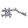
COPPER (II) ION / DINUCLEAR COPPER ION / HEME-AS / PROTOPORPHYRIN IX CONTAINING FE / (2R)-2,3-dihydroxypropyl (9Z)-octadec-9-enoate / Cytochrome c oxidase polypeptide 2A / Cytochrome c oxidase subunit 1 / Cytochrome c oxidase subunit 2
Similarity search - Component
Biological speciesThermus thermophilus (bacteria)
MethodX-RAY DIFFRACTION / SYNCHROTRON / MOLECULAR REPLACEMENT / Resolution: 2.28 Å
AuthorsKabbinale, A. / Johannesson, J. / Finke, D.
Funding supportEuropean Union, Sweden, 4items
OrganizationGrant numberCountry
European Research Council (ERC)789030European Union
European Research Council (ERC)963936European Union
Swedish Research Council2021-05662 Sweden
Swedish Research Council2015-00560 Sweden
CitationJournal: To Be Published
Title: Structural changes in cytochrome c oxidase following the reduction of dioxygen to water
Authors: Zoric, D. / Johannesson, J. / Kabbinale, A. / Sandelin, E. / Vallejos, A. / Ghosh, S. / Dahl, P. / Ronnholm, J. / Bjelcic, M. / Finke, A. / Bostedt, C. / Bacellar Cases da Silveira, C. / ...Authors: Zoric, D. / Johannesson, J. / Kabbinale, A. / Sandelin, E. / Vallejos, A. / Ghosh, S. / Dahl, P. / Ronnholm, J. / Bjelcic, M. / Finke, A. / Bostedt, C. / Bacellar Cases da Silveira, C. / Beale, E. / Cirelli, C. / Johnson, P. / Ozerov, D. / Boutet, S. / Batyuk, A. / Kupitz, C. / Peck, A. / Poitevin, F. / Sierra, R. / Lisova, S. / Wallentin, C. / Branden, G. / Ostojic, L. / Glerup, J. / Neutze, R.
History
DepositionAug 5, 2024Deposition site: PDBE / Processing site: PDBE
Revision 1.0Aug 13, 2025Provider: repository / Type: Initial release

-
Structure visualization

Structure viewerMolecule:
MolmilJmol/JSmol

Downloads & links

-
Assembly

Deposited unit
A: Cytochrome c oxidase subunit 1
B: Cytochrome c oxidase subunit 2
C: Cytochrome c oxidase polypeptide 2A
hetero molecules


Theoretical massNumber of molelcules
Total (without water)93,03824
Polymers85,8913
Non-polymers7,14721
Water1,910106
1


  • Idetical with deposited unit
  • defined by author
  • Evidence: gel filtration
TypeNameSymmetry operationNumber
identity operation1_555x,y,z1
Unit cell
Length a, b, c (Å)143.688, 98.340, 94.085
Angle α, β, γ (deg.)90.00, 127.58, 90.00
Int Tables number5
Space group name H-MC121

-
Components

-
Cytochrome c oxidase subunit ... , 2 types, 2 molecules AB

#1: Protein Cytochrome c oxidase subunit 1 / Cytochrome c ba(3) subunit I / Cytochrome c oxidase polypeptide I / Cytochrome cba3 subunit 1


Mass: 63540.141 Da / Num. of mol.: 1
Source method: isolated from a genetically manipulated source
Source: (gene. exp.) Thermus thermophilus (bacteria) / Gene: cbaA, TTHA1135 / Production host: Thermus thermophilus HB8 (bacteria) / Strain (production host): DSM 579 / HB8 / References: UniProt: Q5SJ79, cytochrome-c oxidase
#2: Protein Cytochrome c oxidase subunit 2 / Cytochrome c ba(3) subunit II / Cytochrome c oxidase polypeptide II / Cytochrome cba3 subunit 2


Mass: 18581.299 Da / Num. of mol.: 1
Source method: isolated from a genetically manipulated source
Source: (gene. exp.) Thermus thermophilus (bacteria) / Gene: cbaB, ctaC, TTHA1134 / Production host: Thermus thermophilus HB8 (bacteria) / Strain (production host): DSM 579 / HB8 / References: UniProt: Q5SJ80, cytochrome-c oxidase

-
Protein/peptide , 1 types, 1 molecules C

#3: Protein/peptide Cytochrome c oxidase polypeptide 2A / Cytochrome c ba(3) subunit IIA / Cytochrome c oxidase polypeptide IIA / Cytochrome cba3 subunit 2A


Mass: 3769.602 Da / Num. of mol.: 1
Source method: isolated from a genetically manipulated source
Source: (gene. exp.) Thermus thermophilus (bacteria) / Gene: cbaD, TTHA1133 / Production host: Thermus thermophilus HB8 (bacteria) / Strain (production host): DSM 579 / HB8 / References: UniProt: P82543, cytochrome-c oxidase

-
Non-polymers , 7 types, 127 molecules

#4: Chemical ChemComp-CU / COPPER (II) ION


Mass: 63.546 Da / Num. of mol.: 1 / Source method: obtained synthetically / Formula: Cu / Feature type: SUBJECT OF INVESTIGATION
#5: Chemical ChemComp-HEM / PROTOPORPHYRIN IX CONTAINING FE / HEME


Mass: 616.487 Da / Num. of mol.: 1 / Source method: obtained synthetically / Formula: C34H32FeN4O4 / Feature type: SUBJECT OF INVESTIGATION
#6: Chemical ChemComp-HAS / HEME-AS


Mass: 920.954 Da / Num. of mol.: 1 / Source method: obtained synthetically / Formula: C54H64FeN4O6 / Feature type: SUBJECT OF INVESTIGATION
#7: Chemical
ChemComp-OLC / (2R)-2,3-dihydroxypropyl (9Z)-octadec-9-enoate / 1-Oleoyl-R-glycerol


Mass: 356.540 Da / Num. of mol.: 15 / Source method: obtained synthetically / Formula: C21H40O4
#8: Chemical ChemComp-CL / CHLORIDE ION


Mass: 35.453 Da / Num. of mol.: 2 / Source method: obtained synthetically / Formula: Cl / Feature type: SUBJECT OF INVESTIGATION
#9: Chemical ChemComp-CUA / DINUCLEAR COPPER ION


Mass: 127.092 Da / Num. of mol.: 1 / Source method: obtained synthetically / Formula: Cu2 / Feature type: SUBJECT OF INVESTIGATION
#10: Water ChemComp-HOH / water


Mass: 18.015 Da / Num. of mol.: 106 / Source method: isolated from a natural source / Formula: H2O

-
Details

Has ligand of interestY
Has protein modificationN

-
Experimental details

-
Experiment

ExperimentMethod: X-RAY DIFFRACTION / Number of used crystals: 1

-
Sample preparation

CrystalDensity Matthews: 3.3 Å3/Da / Density % sol: 62.69 %
Crystal growTemperature: 298 K / Method: lipidic cubic phase / pH: 5.3 / Details: 1.4 M NaCl, 100 mM MES. 40-43 % (v/v) PEG 400 / Temp details: Cryogenic

-
Data collection

DiffractionMean temperature: 100 K / Ambient temp details: Cryogenic / Serial crystal experiment: N
Diffraction sourceSource: SYNCHROTRON / Site: MAX IV / Beamline: BioMAX / Wavelength: 1.9997 Å
DetectorType: DECTRIS EIGER X 16M / Detector: PIXEL / Date: Feb 10, 2023 / Details: Kirkpatrick-Baez (KB) mirror pair (VFM, HFM)
RadiationMonochromator: double crystal / Protocol: SINGLE WAVELENGTH / Monochromatic (M) / Laue (L): M / Scattering type: x-ray
Radiation wavelengthWavelength: 1.9997 Å / Relative weight: 1
ReflectionResolution: 2.28→74.54 Å / Num. obs: 44643 / % possible obs: 94.4 % / Redundancy: 9.2 % / CC1/2: 0.998 / Net I/σ(I): 18
Reflection shellResolution: 2.28→2.36 Å / Num. unique obs: 2943 / CC1/2: 0.727

-
Processing

Software
NameVersionClassification
REFMAC5.8.0419refinement
autoPROCdata reduction
STARANISOdata scaling
PHASERphasing
RefinementMethod to determine structure: MOLECULAR REPLACEMENT / Resolution: 2.28→74.54 Å / Cor.coef. Fo:Fc: 0.96 / Cor.coef. Fo:Fc free: 0.927 / SU B: 3.902 / SU ML: 0.098 / Cross valid method: THROUGHOUT / ESU R: 0.223 / ESU R Free: 0.18 / Stereochemistry target values: MAXIMUM LIKELIHOOD / Details: HYDROGENS HAVE BEEN ADDED IN THE RIDING POSITIONS
RfactorNum. reflection% reflectionSelection details
Rfree0.19652 2187 4.9 %RANDOM
Rwork0.15308 ---
obs0.15527 42456 94.34 %-
Solvent computationIon probe radii: 0.8 Å / Shrinkage radii: 0.8 Å / VDW probe radii: 1.2 Å / Solvent model: MASK
Displacement parametersBiso mean: 47.693 Å2
Baniso -1Baniso -2Baniso -3
1-0.5 Å2-0 Å21.59 Å2
2---2.11 Å20 Å2
3----0.38 Å2
Refinement stepCycle: 1 / Resolution: 2.28→74.54 Å
ProteinNucleic acidLigandSolventTotal
Num. atoms5910 0 386 106 6402
Refine LS restraints
Refine-IDTypeDev idealDev ideal targetNumber
X-RAY DIFFRACTIONr_bond_refined_d0.0090.0126506
X-RAY DIFFRACTIONr_bond_other_d0.0010.0166404
X-RAY DIFFRACTIONr_angle_refined_deg1.7041.6738845
X-RAY DIFFRACTIONr_angle_other_deg0.5371.58214648
X-RAY DIFFRACTIONr_dihedral_angle_1_deg6.65749
X-RAY DIFFRACTIONr_dihedral_angle_2_deg17.6377.29248
X-RAY DIFFRACTIONr_dihedral_angle_3_deg16.81410890
X-RAY DIFFRACTIONr_dihedral_angle_4_deg
X-RAY DIFFRACTIONr_chiral_restr0.0810.2969
X-RAY DIFFRACTIONr_gen_planes_refined0.010.027368
X-RAY DIFFRACTIONr_gen_planes_other0.0060.021539
X-RAY DIFFRACTIONr_nbd_refined
X-RAY DIFFRACTIONr_nbd_other
X-RAY DIFFRACTIONr_nbtor_refined
X-RAY DIFFRACTIONr_nbtor_other
X-RAY DIFFRACTIONr_xyhbond_nbd_refined
X-RAY DIFFRACTIONr_xyhbond_nbd_other
X-RAY DIFFRACTIONr_metal_ion_refined
X-RAY DIFFRACTIONr_metal_ion_other
X-RAY DIFFRACTIONr_symmetry_vdw_refined
X-RAY DIFFRACTIONr_symmetry_vdw_other
X-RAY DIFFRACTIONr_symmetry_hbond_refined
X-RAY DIFFRACTIONr_symmetry_hbond_other
X-RAY DIFFRACTIONr_symmetry_metal_ion_refined
X-RAY DIFFRACTIONr_symmetry_metal_ion_other
X-RAY DIFFRACTIONr_mcbond_it3.944.5153005
X-RAY DIFFRACTIONr_mcbond_other3.9394.5153004
X-RAY DIFFRACTIONr_mcangle_it5.1348.1123751
X-RAY DIFFRACTIONr_mcangle_other5.1358.1113752
X-RAY DIFFRACTIONr_scbond_it5.2885.1973501
X-RAY DIFFRACTIONr_scbond_other5.2875.1993502
X-RAY DIFFRACTIONr_scangle_it
X-RAY DIFFRACTIONr_scangle_other7.529.2435095
X-RAY DIFFRACTIONr_long_range_B_refined8.51244.557299
X-RAY DIFFRACTIONr_long_range_B_other8.51244.557299
X-RAY DIFFRACTIONr_rigid_bond_restr
X-RAY DIFFRACTIONr_sphericity_free
X-RAY DIFFRACTIONr_sphericity_bonded
LS refinement shellResolution: 2.281→2.34 Å / Total num. of bins used: 20
RfactorNum. reflection% reflection
Rfree0.224 92 -
Rwork0.179 2119 -
obs--64.03 %

+
About Yorodumi

-
News

-
Feb 9, 2022. New format data for meta-information of EMDB entries

New format data for meta-information of EMDB entries

  • Version 3 of the EMDB header file is now the official format.
  • The previous official version 1.9 will be removed from the archive.

Related info.:EMDB header

External links:wwPDB to switch to version 3 of the EMDB data model

-
Aug 12, 2020. Covid-19 info

Covid-19 info

URL: https://pdbj.org/emnavi/covid19.php

New page: Covid-19 featured information page in EM Navigator.

Related info.:Covid-19 info / Mar 5, 2020. Novel coronavirus structure data

+
Mar 5, 2020. Novel coronavirus structure data

Novel coronavirus structure data

Related info.:Yorodumi Speices / Aug 12, 2020. Covid-19 info

External links:COVID-19 featured content - PDBj / Molecule of the Month (242):Coronavirus Proteases

+
Jan 31, 2019. EMDB accession codes are about to change! (news from PDBe EMDB page)

EMDB accession codes are about to change! (news from PDBe EMDB page)

  • The allocation of 4 digits for EMDB accession codes will soon come to an end. Whilst these codes will remain in use, new EMDB accession codes will include an additional digit and will expand incrementally as the available range of codes is exhausted. The current 4-digit format prefixed with “EMD-” (i.e. EMD-XXXX) will advance to a 5-digit format (i.e. EMD-XXXXX), and so on. It is currently estimated that the 4-digit codes will be depleted around Spring 2019, at which point the 5-digit format will come into force.
  • The EM Navigator/Yorodumi systems omit the EMD- prefix.

Related info.:Q: What is EMD? / ID/Accession-code notation in Yorodumi/EM Navigator

External links:EMDB Accession Codes are Changing Soon! / Contact to PDBj

+
Jul 12, 2017. Major update of PDB

Major update of PDB

  • wwPDB released updated PDB data conforming to the new PDBx/mmCIF dictionary.
  • This is a major update changing the version number from 4 to 5, and with Remediation, in which all the entries are updated.
  • In this update, many items about electron microscopy experimental information are reorganized (e.g. em_software).
  • Now, EM Navigator and Yorodumi are based on the updated data.

External links:wwPDB Remediation / Enriched Model Files Conforming to OneDep Data Standards Now Available in the PDB FTP Archive

-
Yorodumi

Thousand views of thousand structures

  • Yorodumi is a browser for structure data from EMDB, PDB, SASBDB, etc.
  • This page is also the successor to EM Navigator detail page, and also detail information page/front-end page for Omokage search.
  • The word "yorodu" (or yorozu) is an old Japanese word meaning "ten thousand". "mi" (miru) is to see.

Related info.:EMDB / PDB / SASBDB / Comparison of 3 databanks / Yorodumi Search / Aug 31, 2016. New EM Navigator & Yorodumi / Yorodumi Papers / Jmol/JSmol / Function and homology information / Changes in new EM Navigator and Yorodumi

Read more