[English] 日本語
 Yorodumi
- PDB-9gcj: The crystal structure of beta-glucosidase from the thermophilic b... -
+ Open data
- Basic information
| Entry | Database: PDB / ID: 9gcj | |||||||||||||||
|---|---|---|---|---|---|---|---|---|---|---|---|---|---|---|---|---|
| Title | The crystal structure of beta-glucosidase from the thermophilic bacterium Caldicellulosiruptor saccharolyticus in complex with beta-D-glucose determined at 1.95 A resolution | |||||||||||||||
 Components Components | beta-glucosidase | |||||||||||||||
 Keywords Keywords | HYDROLASE / Biocatalysis / beta-glucosidase / Caldicellulosiruptor saccharolyticus | |||||||||||||||
| Function / homology |  Function and homology information Function and homology informationbeta-glucosidase / beta-glucosidase activity / cellulose catabolic process / cytosol Similarity search - Function | |||||||||||||||
| Biological species |   Caldicellulosiruptor saccharolyticus DSM 8903 (bacteria) Caldicellulosiruptor saccharolyticus DSM 8903 (bacteria) | |||||||||||||||
| Method |  X-RAY DIFFRACTION / X-RAY DIFFRACTION /  SYNCHROTRON / SYNCHROTRON /  MOLECULAR REPLACEMENT / Resolution: 1.95 Å MOLECULAR REPLACEMENT / Resolution: 1.95 Å | |||||||||||||||
 Authors Authors | Chrysina, E.D. / Sotiropoulou, A.I. | |||||||||||||||
| Funding support | European Union, 4items
| |||||||||||||||
 Citation Citation |  Journal: Acta Crystallogr D Struct Biol / Year: 2024 Journal: Acta Crystallogr D Struct Biol / Year: 2024Title: Structural studies of beta-glucosidase from the thermophilic bacterium Caldicellulosiruptor saccharolyticus. Authors: Sotiropoulou, A.I. / Hatzinikolaou, D.G. / Chrysina, E.D. | |||||||||||||||
| History |
|
- Structure visualization
| Structure viewer | Molecule:  Molmil Molmil Jmol/JSmol Jmol/JSmol |
|---|
- Downloads & links
- Download
| PDBx/mmCIF format |  9gcj.cif.gz 9gcj.cif.gz | 382.2 KB | Display |  PDBx/mmCIF format PDBx/mmCIF format |
|---|---|---|---|---|
| PDB format |  pdb9gcj.ent.gz pdb9gcj.ent.gz | 303.7 KB | Display |  PDB format PDB format |
| PDBx/mmJSON format |  9gcj.json.gz 9gcj.json.gz | Tree view |  PDBx/mmJSON format PDBx/mmJSON format | |
| Others |  Other downloads Other downloads |
-Validation report
| Arichive directory |  https://data.pdbj.org/pub/pdb/validation_reports/gc/9gcj https://data.pdbj.org/pub/pdb/validation_reports/gc/9gcj ftp://data.pdbj.org/pub/pdb/validation_reports/gc/9gcj ftp://data.pdbj.org/pub/pdb/validation_reports/gc/9gcj | HTTPS FTP |
|---|
-Related structure data
| Related structure data |  9gciC C: citing same article ( |
|---|---|
| Similar structure data | Similarity search - Function & homology  F&H Search F&H Search |
- Links
Links
- Assembly
| Deposited unit | 
| ||||||||
|---|---|---|---|---|---|---|---|---|---|
| 1 | 
| ||||||||
| Unit cell |
|
- Components
-Protein / Sugars , 2 types, 4 molecules AB

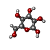
| #1: Protein | Mass: 54175.371 Da / Num. of mol.: 2 Source method: isolated from a genetically manipulated source Details: The actual sequence of the enzyme is deposited with Uniprotkb with entry code: A4XIG7_CALS8 (https://www.uniprot.org/uniprotkb/A4XIG7/entry). In the present structure, a total of five (5) ...Details: The actual sequence of the enzyme is deposited with Uniprotkb with entry code: A4XIG7_CALS8 (https://www.uniprot.org/uniprotkb/A4XIG7/entry). In the present structure, a total of five (5) amino acids were modelled at the N-terminus of Bgl1:molA. These residues originate from the translated sequence of the pET15b plasmid section (eight (8) amino acids, GSHMLEDP) between the thrombin cleavage site and the BamH I site in the multicloning area of the plasmid. Source: (gene. exp.)   Caldicellulosiruptor saccharolyticus DSM 8903 (bacteria) Caldicellulosiruptor saccharolyticus DSM 8903 (bacteria)Gene: Csac_1089 Production host:  References: UniProt: A4XIG7, beta-glucosidase #2: Sugar | |
|---|
-Non-polymers , 4 types, 406 molecules 


| #3: Chemical | | #4: Chemical | ChemComp-PEG / | #5: Chemical | ChemComp-CL / | #6: Water | ChemComp-HOH / | |
|---|
-Details
| Has ligand of interest | Y |
|---|---|
| Has protein modification | N |
| Nonpolymer details | Additional portions of density have been observed in the structure that could not be attributed to ...Additional portions of density have been observed in the structure that could not be attributed to any of the known molecules that were present during sample preparation, crystallisation and X-ray data collection. Non-specific binding of polyethylene glycol molecules is observed but there was not sufficient density to include all of them in the structure. These molecules have not been correlated with the functional role of the enzyme. |
-Experimental details
-Experiment
| Experiment | Method:  X-RAY DIFFRACTION / Number of used crystals: 1 X-RAY DIFFRACTION / Number of used crystals: 1 |
|---|
- Sample preparation
| Crystal | Density Matthews: 2.52 Å3/Da / Density % sol: 51.18 % / Description: Thin plates |
|---|---|
| Crystal grow | Temperature: 292.15 K / Method: vapor diffusion, sitting drop / pH: 5.5 Details: Co-crystals: 0.2 M magnesium chloride hexahydrate, 0.1M Bis-Tris, pH 5.5, 25% (w/v) PEG 3350 in the presence of 0.6 mM lactose |
-Data collection
| Diffraction | Mean temperature: 100 K / Serial crystal experiment: N |
|---|---|
| Diffraction source | Source:  SYNCHROTRON / Site: SYNCHROTRON / Site:  PETRA III, EMBL c/o DESY PETRA III, EMBL c/o DESY  / Beamline: P13 (MX1) / Wavelength: 0.9763 Å / Beamline: P13 (MX1) / Wavelength: 0.9763 Å |
| Detector | Type: DECTRIS PILATUS 12M / Detector: PIXEL / Date: Jul 8, 2021 |
| Radiation | Monochromator: SI(111) / Protocol: SINGLE WAVELENGTH / Monochromatic (M) / Laue (L): M / Scattering type: x-ray |
| Radiation wavelength | Wavelength: 0.9763 Å / Relative weight: 1 |
| Reflection | Resolution: 1.95→98.31 Å / Num. obs: 77506 / % possible obs: 99.6 % / Redundancy: 6.7 % / CC1/2: 0.974 / Rpim(I) all: 0.095 / Rrim(I) all: 0.137 / Net I/σ(I): 8.3 |
| Reflection shell | Resolution: 1.95→1.99 Å / Redundancy: 6.7 % / Mean I/σ(I) obs: 1.8 / Num. unique obs: 4600 / CC1/2: 0.653 / Rpim(I) all: 0.634 / Rrim(I) all: 1.655 / % possible all: 99.9 |
- Processing
| Software |
| |||||||||||||||||||||||||||||||||||||||||||||||||||||||||||||||||||||||||||||||||||||||||||||||||||||||||||||||||||||||||||||||||||||||||||||||||||||||||||||||||||||||||||||||||||||||||||||||||||||||||||||||||||||||||||||||||||||||
|---|---|---|---|---|---|---|---|---|---|---|---|---|---|---|---|---|---|---|---|---|---|---|---|---|---|---|---|---|---|---|---|---|---|---|---|---|---|---|---|---|---|---|---|---|---|---|---|---|---|---|---|---|---|---|---|---|---|---|---|---|---|---|---|---|---|---|---|---|---|---|---|---|---|---|---|---|---|---|---|---|---|---|---|---|---|---|---|---|---|---|---|---|---|---|---|---|---|---|---|---|---|---|---|---|---|---|---|---|---|---|---|---|---|---|---|---|---|---|---|---|---|---|---|---|---|---|---|---|---|---|---|---|---|---|---|---|---|---|---|---|---|---|---|---|---|---|---|---|---|---|---|---|---|---|---|---|---|---|---|---|---|---|---|---|---|---|---|---|---|---|---|---|---|---|---|---|---|---|---|---|---|---|---|---|---|---|---|---|---|---|---|---|---|---|---|---|---|---|---|---|---|---|---|---|---|---|---|---|---|---|---|---|---|---|---|---|---|---|---|---|---|---|---|---|---|---|---|---|---|---|---|---|
| Refinement | Method to determine structure:  MOLECULAR REPLACEMENT / Resolution: 1.95→62.615 Å / Cor.coef. Fo:Fc: 0.963 / Cor.coef. Fo:Fc free: 0.951 / SU B: 4.161 / SU ML: 0.111 / Cross valid method: FREE R-VALUE / ESU R: 0.15 / ESU R Free: 0.136 MOLECULAR REPLACEMENT / Resolution: 1.95→62.615 Å / Cor.coef. Fo:Fc: 0.963 / Cor.coef. Fo:Fc free: 0.951 / SU B: 4.161 / SU ML: 0.111 / Cross valid method: FREE R-VALUE / ESU R: 0.15 / ESU R Free: 0.136 Details: Hydrogens have been added in their riding positions
| |||||||||||||||||||||||||||||||||||||||||||||||||||||||||||||||||||||||||||||||||||||||||||||||||||||||||||||||||||||||||||||||||||||||||||||||||||||||||||||||||||||||||||||||||||||||||||||||||||||||||||||||||||||||||||||||||||||||
| Solvent computation | Ion probe radii: 0.8 Å / Shrinkage radii: 0.8 Å / VDW probe radii: 1.2 Å / Solvent model: MASK BULK SOLVENT | |||||||||||||||||||||||||||||||||||||||||||||||||||||||||||||||||||||||||||||||||||||||||||||||||||||||||||||||||||||||||||||||||||||||||||||||||||||||||||||||||||||||||||||||||||||||||||||||||||||||||||||||||||||||||||||||||||||||
| Displacement parameters | Biso mean: 31.517 Å2
| |||||||||||||||||||||||||||||||||||||||||||||||||||||||||||||||||||||||||||||||||||||||||||||||||||||||||||||||||||||||||||||||||||||||||||||||||||||||||||||||||||||||||||||||||||||||||||||||||||||||||||||||||||||||||||||||||||||||
| Refinement step | Cycle: LAST / Resolution: 1.95→62.615 Å
| |||||||||||||||||||||||||||||||||||||||||||||||||||||||||||||||||||||||||||||||||||||||||||||||||||||||||||||||||||||||||||||||||||||||||||||||||||||||||||||||||||||||||||||||||||||||||||||||||||||||||||||||||||||||||||||||||||||||
| Refine LS restraints |
| |||||||||||||||||||||||||||||||||||||||||||||||||||||||||||||||||||||||||||||||||||||||||||||||||||||||||||||||||||||||||||||||||||||||||||||||||||||||||||||||||||||||||||||||||||||||||||||||||||||||||||||||||||||||||||||||||||||||
| LS refinement shell | Refine-ID: X-RAY DIFFRACTION / Total num. of bins used: 20
|
 Movie
Movie Controller


 PDBj
PDBj


