+ Open data
- Basic information
| Entry | Database: PDB / ID: 9gb7 | ||||||||||||||||||||||||
|---|---|---|---|---|---|---|---|---|---|---|---|---|---|---|---|---|---|---|---|---|---|---|---|---|---|
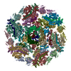
| Title | Extended phiCD508 neck | ||||||||||||||||||||||||
 Components Components |
| ||||||||||||||||||||||||
 Keywords Keywords | VIRUS / Bacteriophage / needle / baseplate | ||||||||||||||||||||||||
| Function / homology |  Function and homology information Function and homology informationPortal protein / Phage portal protein, SPP1 Gp6-like / Phage tail tube protein / XkdM-like superfamily / Phage tail tube protein / Phage tail sheath protein, beta-sandwich domain / Phage tail sheath protein beta-sandwich domain / Tail sheath protein, subtilisin-like domain / Phage tail sheath protein subtilisin-like domain / Tail sheath protein, C-terminal domain / Phage tail sheath C-terminal domain Similarity search - Domain/homology | ||||||||||||||||||||||||
| Biological species |  Clostridioides difficile (bacteria) Clostridioides difficile (bacteria) | ||||||||||||||||||||||||
| Method | ELECTRON MICROSCOPY / single particle reconstruction / cryo EM / Resolution: 3.4 Å | ||||||||||||||||||||||||
 Authors Authors | Wilson, J.S. / Fagan, R.P. / Bullough, P.A. | ||||||||||||||||||||||||
| Funding support |  United Kingdom, 1items United Kingdom, 1items
| ||||||||||||||||||||||||
 Citation Citation |  Journal: Life Sci Alliance / Year: 2025 Journal: Life Sci Alliance / Year: 2025Title: Molecular mechanism of bacteriophage contraction structure of an S-layer-penetrating bacteriophage. Authors: Jason S Wilson / Louis-Charles Fortier / Robert P Fagan / Per A Bullough /   Abstract: The molecular details of phage tail contraction and bacterial cell envelope penetration remain poorly understood and are completely unknown for phages infecting bacteria enveloped by proteinaceous S- ...The molecular details of phage tail contraction and bacterial cell envelope penetration remain poorly understood and are completely unknown for phages infecting bacteria enveloped by proteinaceous S-layers. Here, we reveal the extended and contracted atomic structures of an intact contractile-tailed phage (φCD508) that binds to and penetrates the protective S-layer of the Gram-positive human pathogen The tail is unusually long (225 nm), and it is also notable that the tail contracts less than those studied in related contractile injection systems such as the model phage T4 (∼20% compared with ∼50%). Surprisingly, we find no evidence of auxiliary enzymatic domains that other phages exploit in cell wall penetration, suggesting that sufficient energy is released upon tail contraction to penetrate the S-layer and the thick cell wall without enzymatic activity. Instead, the unusually long tail length, which becomes more flexible upon contraction, likely contributes toward the required free energy release for envelope penetration. | ||||||||||||||||||||||||
| History |
|
- Structure visualization
| Structure viewer | Molecule:  Molmil Molmil Jmol/JSmol Jmol/JSmol |
|---|
- Downloads & links
- Download
| PDBx/mmCIF format |  9gb7.cif.gz 9gb7.cif.gz | 4.3 MB | Display |  PDBx/mmCIF format PDBx/mmCIF format |
|---|---|---|---|---|
| PDB format |  pdb9gb7.ent.gz pdb9gb7.ent.gz | Display |  PDB format PDB format | |
| PDBx/mmJSON format |  9gb7.json.gz 9gb7.json.gz | Tree view |  PDBx/mmJSON format PDBx/mmJSON format | |
| Others |  Other downloads Other downloads |
-Validation report
| Summary document |  9gb7_validation.pdf.gz 9gb7_validation.pdf.gz | 1.7 MB | Display |  wwPDB validaton report wwPDB validaton report |
|---|---|---|---|---|
| Full document |  9gb7_full_validation.pdf.gz 9gb7_full_validation.pdf.gz | 1.7 MB | Display | |
| Data in XML |  9gb7_validation.xml.gz 9gb7_validation.xml.gz | 264.6 KB | Display | |
| Data in CIF |  9gb7_validation.cif.gz 9gb7_validation.cif.gz | 425 KB | Display | |
| Arichive directory |  https://data.pdbj.org/pub/pdb/validation_reports/gb/9gb7 https://data.pdbj.org/pub/pdb/validation_reports/gb/9gb7 ftp://data.pdbj.org/pub/pdb/validation_reports/gb/9gb7 ftp://data.pdbj.org/pub/pdb/validation_reports/gb/9gb7 | HTTPS FTP |
-Related structure data
| Related structure data |  51200MC  9g8sC  9gayC  9gazC  9gb0C  9gb1C  9gb2C  9gb3C  9gb4C  9gb5C  9gb6C  9gb8C M: map data used to model this data C: citing same article ( |
|---|---|
| Similar structure data | Similarity search - Function & homology  F&H Search F&H Search |
- Links
Links
- Assembly
| Deposited unit | 
|
|---|---|
| 1 |
|
- Components
-Protein , 6 types, 48 molecules ABCDEFGKMOQXHLNPRdIJSTUVWYZabc...
| #1: Protein | Mass: 15572.538 Da / Num. of mol.: 6 / Source method: isolated from a natural source / Source: (natural)  Clostridioides difficile (bacteria) / Strain: CD117 / References: UniProt: A0A9X8RMX9 Clostridioides difficile (bacteria) / Strain: CD117 / References: UniProt: A0A9X8RMX9#2: Protein | Mass: 14433.568 Da / Num. of mol.: 6 / Source method: isolated from a natural source / Source: (natural)  Clostridioides difficile (bacteria) / Strain: CD117 / References: UniProt: A0A9X8WSH7 Clostridioides difficile (bacteria) / Strain: CD117 / References: UniProt: A0A9X8WSH7#3: Protein | Mass: 31761.447 Da / Num. of mol.: 6 / Source method: isolated from a natural source / Source: (natural)  Clostridioides difficile (bacteria) / Strain: CD117 / References: UniProt: A0A9X8WSH1 Clostridioides difficile (bacteria) / Strain: CD117 / References: UniProt: A0A9X8WSH1#4: Protein | Mass: 12932.699 Da / Num. of mol.: 12 / Source method: isolated from a natural source / Source: (natural)  Clostridioides difficile (bacteria) / Strain: CD117 / References: UniProt: A0A9X8WSI0 Clostridioides difficile (bacteria) / Strain: CD117 / References: UniProt: A0A9X8WSI0#5: Protein | Mass: 53016.762 Da / Num. of mol.: 6 / Source method: isolated from a natural source / Source: (natural)  Clostridioides difficile (bacteria) / Strain: CD117 / References: UniProt: A0A9X8RMY4 Clostridioides difficile (bacteria) / Strain: CD117 / References: UniProt: A0A9X8RMY4#6: Protein | Mass: 57239.852 Da / Num. of mol.: 12 / Source method: isolated from a natural source / Source: (natural)  Clostridioides difficile (bacteria) / Strain: CD117 / References: UniProt: A0A069A478 Clostridioides difficile (bacteria) / Strain: CD117 / References: UniProt: A0A069A478 |
|---|
-Details
| Has protein modification | N |
|---|
-Experimental details
-Experiment
| Experiment | Method: ELECTRON MICROSCOPY |
|---|---|
| EM experiment | Aggregation state: PARTICLE / 3D reconstruction method: single particle reconstruction |
- Sample preparation
| Component | Name: Extended phiCD508 neck / Type: COMPLEX / Entity ID: all / Source: NATURAL |
|---|---|
| Source (natural) | Organism:  Clostridioides difficile (bacteria) / Strain: CD117 Clostridioides difficile (bacteria) / Strain: CD117 |
| Buffer solution | pH: 7.4 |
| Specimen | Embedding applied: NO / Shadowing applied: NO / Staining applied: NO / Vitrification applied: YES |
| Vitrification | Cryogen name: ETHANE |
- Electron microscopy imaging
| Experimental equipment |  Model: Titan Krios / Image courtesy: FEI Company |
|---|---|
| Microscopy | Model: TFS KRIOS |
| Electron gun | Electron source:  FIELD EMISSION GUN / Accelerating voltage: 300 kV / Illumination mode: FLOOD BEAM FIELD EMISSION GUN / Accelerating voltage: 300 kV / Illumination mode: FLOOD BEAM |
| Electron lens | Mode: BRIGHT FIELD / Nominal defocus max: 3000 nm / Nominal defocus min: 1500 nm |
| Image recording | Electron dose: 42 e/Å2 / Film or detector model: GATAN K3 (6k x 4k) |
- Processing
| CTF correction | Type: PHASE FLIPPING AND AMPLITUDE CORRECTION |
|---|---|
| 3D reconstruction | Resolution: 3.4 Å / Resolution method: FSC 0.143 CUT-OFF / Num. of particles: 23724 / Symmetry type: POINT |
 Movie
Movie Controller


 PDBj
PDBj