Movie
Movie Controller

[English] 日本語
 Yorodumi
- PDB-9g9x: Structure of the human two pore domain potassium ion channel TASK... -
+ Open data

- Basic information
| Entry | Database: PDB / ID: 9g9x | ||||||||||||
|---|---|---|---|---|---|---|---|---|---|---|---|---|---|
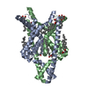
| Title | Structure of the human two pore domain potassium ion channel TASK-1 (K2P3.1) | ||||||||||||
 Components Components | Potassium channel subfamily K member 3 | ||||||||||||
 Keywords Keywords | MEMBRANE PROTEIN / K2P / potassium channel / ion channel | ||||||||||||
| Function / homology |  Function and homology information Function and homology informationopen rectifier potassium channel activity / TWIK-releated acid-sensitive K+ channel (TASK) / regulation of action potential firing rate / detection of hypoxic conditions in blood by carotid body chemoreceptor signaling / Phase 4 - resting membrane potential / potassium ion leak channel activity / regulation of resting membrane potential / S100 protein binding / outward rectifier potassium channel activity / negative regulation of cytosolic calcium ion concentration ...open rectifier potassium channel activity / TWIK-releated acid-sensitive K+ channel (TASK) / regulation of action potential firing rate / detection of hypoxic conditions in blood by carotid body chemoreceptor signaling / Phase 4 - resting membrane potential / potassium ion leak channel activity / regulation of resting membrane potential / S100 protein binding / outward rectifier potassium channel activity / negative regulation of cytosolic calcium ion concentration / sodium channel activity / cellular response to zinc ion / cochlea development / monoatomic ion channel activity / potassium channel activity / cellular response to acidic pH / potassium ion transmembrane transport / potassium ion transport / monoatomic ion transmembrane transport / cellular response to hypoxia / chemical synaptic transmission / response to xenobiotic stimulus / protein heterodimerization activity / synapse / metal ion binding / plasma membrane Similarity search - Function | ||||||||||||
| Biological species |  Homo sapiens (human) Homo sapiens (human) | ||||||||||||
| Method | ELECTRON MICROSCOPY / single particle reconstruction / cryo EM / Resolution: 3.13 Å | ||||||||||||
 Authors Authors | Rodstrom, K.E.J. / Hall, P.H. / Tucker, S.J. | ||||||||||||
| Funding support |  United Kingdom, 3items United Kingdom, 3items
| ||||||||||||
 Citation Citation | Journal: Structure / Year: 2025 Title: Structures of TASK-1 and TASK-3 K2P channels provide insight into their gating and dysfunction in disease. Authors: Peter Rory Hall / Thibault Jouen-Tachoire / Marcus Schewe / Peter Proks / Thomas Baukrowitz / Elisabeth P Carpenter / Simon Newstead / Karin E J Rödström / Stephen J Tucker /  Abstract: TASK-1 and TASK-3 are pH-sensitive two-pore domain (K2P/KCNK) K channels. Their functional roles make them promising targets for treatment of multiple disorders including sleep apnea, pain, and ...TASK-1 and TASK-3 are pH-sensitive two-pore domain (K2P/KCNK) K channels. Their functional roles make them promising targets for treatment of multiple disorders including sleep apnea, pain, and atrial fibrillation. Mutations in these channels are also associated with neurodevelopmental and hypertensive disorders. A previous crystal structure of TASK-1 revealed a lower "X-gate" as a hotspot for missense gain-of-function (GoF) mutations associated with DDSA (developmental delay with sleep apnea). However, the mechanisms of gating in TASK channels are still not fully understood. Here, we resolve structures for both human TASK-1 and TASK-3 by cryoelectron microscopy (cryo-EM), as well as a recurrent TASK-3 variant (G236R) associated with KCNK9 imprinting syndrome (KIS) (formerly known as Birk-Barel syndrome). Combined with functional studies of the X-gating mechanism, we provide evidence for how a highly conserved gating mechanism becomes defective in disease, and also provide further insight into the pathway of conformational changes that underlie the pH-dependent inhibition of TASK channel activity. | ||||||||||||
| History |
|
- Structure visualization
| Structure viewer | Molecule: MolmilJmol/JSmol |
|---|
- Downloads & links
-Download
| PDBx/mmCIF format | 9g9x.cif.gz | 110.5 KB | Display | PDBx/mmCIF format |
|---|---|---|---|---|
| PDB format | pdb9g9x.ent.gz | 83.8 KB | Display | PDB format |
| PDBx/mmJSON format | 9g9x.json.gz | Tree view | PDBx/mmJSON format | |
| Others |  Other downloads Other downloads |
-Validation report
| Arichive directory | https://data.pdbj.org/pub/pdb/validation_reports/g9/9g9xftp://data.pdbj.org/pub/pdb/validation_reports/g9/9g9x | HTTPS FTP |
|---|
-Related structure data
| Related structure data |  51160MC  9g9vC 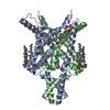 9g9wC M: map data used to model this data C: citing same article ( |
|---|---|
| Similar structure data |

-Links
 PDBj
PDBj

- Assembly
| Deposited unit | 
|
|---|---|
| 1 |
|
-Components
| #1: Protein | Mass: 30117.014 Da / Num. of mol.: 2 Source method: isolated from a genetically manipulated source Details: K2P3.1 residues M1 to E259 with a TEV protease site. Fused purification tags were cleaved prior to EM sample preparation. Source: (gene. exp.) Homo sapiens (human) / Gene: KCNK3, TASK, TASK1 / Plasmid: pFB-CT10HF-LIC / Cell line (production host): Sf9 / Production host:   Spodoptera frugiperda (fall armyworm) / References: UniProt: O14649 Spodoptera frugiperda (fall armyworm) / References: UniProt: O14649#2: Chemical | ChemComp-Y01 /   Mass: 486.726 Da / Num. of mol.: 4 / Source method: obtained synthetically / Formula: C31H50O4 Mass: 486.726 Da / Num. of mol.: 4 / Source method: obtained synthetically / Formula: C31H50O4#3: Chemical |   Mass: 39.098 Da / Num. of mol.: 3 / Source method: obtained synthetically / Formula: K Mass: 39.098 Da / Num. of mol.: 3 / Source method: obtained synthetically / Formula: KHas ligand of interest | N | Has protein modification | N | |
|---|
-Experimental details
-Experiment
| Experiment | Method: ELECTRON MICROSCOPY |
|---|---|
| EM experiment | Aggregation state: PARTICLE / 3D reconstruction method: single particle reconstruction |
- Sample preparation
| Component | Name: K2P3.1 homodimer / Type: COMPLEX Details: Protein generated by removal of the 10xHis and FLAG purification tags with TEV protease cleavage Entity ID: #1 / Source: RECOMBINANT |
|---|---|
| Molecular weight | Value: 0.60 MDa / Experimental value: NO |
| Source (natural) | Organism: Homo sapiens (human) |
| Source (recombinant) | Organism: Spodoptera frugiperda (fall armyworm) / Strain: Sf9 |
| Buffer solution | pH: 7.5 Details: 20 mM HEPES pH 7.5, 200 mM KCl, 0.045% w/v UDM, 0.0045% w/v CHS |
| Specimen | Conc.: 3.6 mg/ml / Embedding applied: NO / Shadowing applied: NO / Staining applied: NO / Vitrification applied: YES / Details: Monodisperse sample |
| Specimen support | Grid material: COPPER / Grid mesh size: 300 divisions/in. / Grid type: Quantifoil R1.2/1.3 |
| Vitrification | Instrument: FEI VITROBOT MARK IV / Cryogen name: ETHANE / Humidity: 100 % / Chamber temperature: 277 K / Details: Grid blotted for 3.5 seconds |
- Electron microscopy imaging
| Experimental equipment |  Model: Titan Krios / Image courtesy: FEI Company |
|---|---|
| Microscopy | Model: FEI TITAN KRIOS |
| Electron gun | Electron source:  FIELD EMISSION GUN / Accelerating voltage: 300 kV / Illumination mode: FLOOD BEAM FIELD EMISSION GUN / Accelerating voltage: 300 kV / Illumination mode: FLOOD BEAM |
| Electron lens | Mode: BRIGHT FIELD / Nominal magnification: 105000 X / Nominal defocus max: 2500 nm / Nominal defocus min: 1250 nm / Alignment procedure: COMA FREE |
| Specimen holder | Cryogen: NITROGEN / Specimen holder model: FEI TITAN KRIOS AUTOGRID HOLDER |
| Image recording | Electron dose: 41.17 e/Å2 / Film or detector model: GATAN K3 BIOQUANTUM (6k x 4k) / Num. of grids imaged: 1 / Num. of real images: 17288 |
| EM imaging optics | Energyfilter name: GIF Bioquantum / Energyfilter slit width: 20 eV |
- Processing
| EM software |
| ||||||||||||||||||||||||||||||||||||
|---|---|---|---|---|---|---|---|---|---|---|---|---|---|---|---|---|---|---|---|---|---|---|---|---|---|---|---|---|---|---|---|---|---|---|---|---|---|
| CTF correction | Type: PHASE FLIPPING AND AMPLITUDE CORRECTION | ||||||||||||||||||||||||||||||||||||
| Symmetry | Point symmetry: C2 (2 fold cyclic) | ||||||||||||||||||||||||||||||||||||
| 3D reconstruction | Resolution: 3.13 Å / Resolution method: FSC 0.143 CUT-OFF / Num. of particles: 117627 / Symmetry type: POINT | ||||||||||||||||||||||||||||||||||||
| Atomic model building | Protocol: FLEXIBLE FIT / Space: REAL | ||||||||||||||||||||||||||||||||||||
| Atomic model building | PDB-ID: 6RV2 Accession code: 6RV2 / Source name: PDB / Type: experimental model |