 Movie
Movie Controller

[English] 日本語
 Yorodumi
- PDB-9g75: Mouse mitochondrial DNA polymerase gamma ternary complex in inter... -
+ Open data

- Basic information
| Entry | Database: PDB / ID: 9g75 | ||||||||||||
|---|---|---|---|---|---|---|---|---|---|---|---|---|---|
| Title | Mouse mitochondrial DNA polymerase gamma ternary complex in intermediate conformer | ||||||||||||
 Components Components |
| ||||||||||||
 Keywords Keywords | TRANSFERASE / Mitochondrial DNA polymerase | ||||||||||||
| Function / homology |  Function and homology information Function and homology informationStrand-asynchronous mitochondrial DNA replication / gamma DNA polymerase complex / mitochondrial chromosome / mitochondrial DNA metabolic process / mitochondrial DNA replication / DNA replication proofreading / single-stranded DNA 3'-5' DNA exonuclease activity / DNA polymerase processivity factor activity / mitochondrial nucleoid / 5'-deoxyribose-5-phosphate lyase activity ...Strand-asynchronous mitochondrial DNA replication / gamma DNA polymerase complex / mitochondrial chromosome / mitochondrial DNA metabolic process / mitochondrial DNA replication / DNA replication proofreading / single-stranded DNA 3'-5' DNA exonuclease activity / DNA polymerase processivity factor activity / mitochondrial nucleoid / 5'-deoxyribose-5-phosphate lyase activity / base-excision repair, gap-filling / DNA polymerase binding / mitochondrion organization / DNA-templated DNA replication / in utero embryonic development / protease binding / double-stranded DNA binding / DNA-directed DNA polymerase / DNA-directed DNA polymerase activity / DNA replication / mitochondrial matrix / DNA repair / chromatin binding / mitochondrion / DNA binding / identical protein binding / cytoplasm Similarity search - Function | ||||||||||||
| Biological species |  synthetic construct (others) | ||||||||||||
| Method | ELECTRON MICROSCOPY / single particle reconstruction / cryo EM / Resolution: 2.98 Å | ||||||||||||
 Authors Authors | Valenzuela, S. / Falkenberg, M. | ||||||||||||
| Funding support |  Sweden, 3items Sweden, 3items
| ||||||||||||
 Citation Citation | Journal: Nat Commun / Year: 2025 Title: Modelling POLG mutations in mice unravels a critical role of POLγΒ in regulating phenotypic severity. Authors: Samantha Corrà / Alessandro Zuppardo / Sebastian Valenzuela / Louise Jenninger / Raffaele Cerutti / Sirelin Sillamaa / Emily Hoberg / Katarina A S Johansson / Urska Rovsnik / Sara Volta / ...Authors: Samantha Corrà / Alessandro Zuppardo / Sebastian Valenzuela / Louise Jenninger / Raffaele Cerutti / Sirelin Sillamaa / Emily Hoberg / Katarina A S Johansson / Urska Rovsnik / Sara Volta / Pedro Silva-Pinheiro / Hannah Davis / Aleksandra Trifunovic / Michal Minczuk / Claes M Gustafsson / Anu Suomalainen / Massimo Zeviani / Bertil Macao / Xuefeng Zhu / Maria Falkenberg / Carlo Viscomi /      Abstract: DNA polymerase γ (POLγ), responsible for mitochondrial DNA replication, consists of a catalytic POLγA subunit and two accessory POLγB subunits. Mutations in POLG, which encodes POLγA, lead to ...DNA polymerase γ (POLγ), responsible for mitochondrial DNA replication, consists of a catalytic POLγA subunit and two accessory POLγB subunits. Mutations in POLG, which encodes POLγA, lead to various mitochondrial diseases. We investigated the most common POLG mutations (A467T, W748S, G848S, Y955C) by characterizing human and mouse POLγ variants. Our data reveal that these mutations significantly impair POLγ activities, with mouse variants exhibiting milder defects. Cryogenic electron microscopy highlighted structural differences between human and mouse POLγ, particularly in the POLγB subunit, which may explain the higher activity of mouse POLγ and the reduced severity of mutations in mice. We further generated a panel of mouse models mirroring common human POLG mutations, providing crucial insights into the pathogenesis of POLG-related disorders and establishing robust models for therapeutic development. Our findings emphasize the importance of POLγB in modulating the severity of POLG mutations. | ||||||||||||
| History |
|
- Structure visualization
| Structure viewer | Molecule: MolmilJmol/JSmol |
|---|
- Downloads & links
-Download
| PDBx/mmCIF format | 9g75.cif.gz | 365.5 KB | Display | PDBx/mmCIF format |
|---|---|---|---|---|
| PDB format | pdb9g75.ent.gz | 280.1 KB | Display | PDB format |
| PDBx/mmJSON format | 9g75.json.gz | Tree view | PDBx/mmJSON format | |
| Others |  Other downloads Other downloads |
-Validation report
| Arichive directory | https://data.pdbj.org/pub/pdb/validation_reports/g7/9g75ftp://data.pdbj.org/pub/pdb/validation_reports/g7/9g75 | HTTPS FTP |
|---|
-Related structure data
| Related structure data |  51110MC  9g74C  9g77C  9ibxC  9ibzC  9ic0C  9ic1C 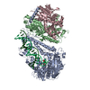 9ic3C M: map data used to model this data C: citing same article ( |
|---|---|
| Similar structure data |


-Links
 PDBj
PDBj


- Assembly
| Deposited unit | 
|
|---|---|
| 1 |
|
-Components
| #1: Protein | Mass: 135078.188 Da / Num. of mol.: 1 Source method: isolated from a genetically manipulated source Source: (gene. exp.)   Spodoptera frugiperda (fall armyworm) / References: UniProt: Q75WC0, DNA-directed DNA polymerase Spodoptera frugiperda (fall armyworm) / References: UniProt: Q75WC0, DNA-directed DNA polymerase | ||||||
|---|---|---|---|---|---|---|---|
| #2: Protein | Mass: 50778.648 Da / Num. of mol.: 2 Source method: isolated from a genetically manipulated source Source: (gene. exp.)  #3: DNA chain | | Mass: 7780.008 Da / Num. of mol.: 1 / Source method: obtained synthetically / Source: (synth.) synthetic construct (others) #4: DNA chain | | Mass: 12162.783 Da / Num. of mol.: 1 / Source method: obtained synthetically / Source: (synth.) synthetic construct (others) Has protein modification | N | |
-Experimental details
-Experiment
| Experiment | Method: ELECTRON MICROSCOPY |
|---|---|
| EM experiment | Aggregation state: PARTICLE / 3D reconstruction method: single particle reconstruction |
- Sample preparation
| Component |
| ||||||||||||||||||||||||
|---|---|---|---|---|---|---|---|---|---|---|---|---|---|---|---|---|---|---|---|---|---|---|---|---|---|
| Source (natural) |
| ||||||||||||||||||||||||
| Source (recombinant) |
| ||||||||||||||||||||||||
| Buffer solution | pH: 7.5 | ||||||||||||||||||||||||
| Specimen | Embedding applied: NO / Shadowing applied: NO / Staining applied: NO / Vitrification applied: YES | ||||||||||||||||||||||||
| Vitrification | Instrument: FEI VITROBOT MARK IV / Cryogen name: ETHANE / Humidity: 100 % |
- Electron microscopy imaging
| Experimental equipment |  Model: Titan Krios / Image courtesy: FEI Company |
|---|---|
| Microscopy | Model: FEI TITAN KRIOS |
| Electron gun | Electron source:  FIELD EMISSION GUN / Accelerating voltage: 300 kV / Illumination mode: FLOOD BEAM FIELD EMISSION GUN / Accelerating voltage: 300 kV / Illumination mode: FLOOD BEAM |
| Electron lens | Mode: BRIGHT FIELD / Nominal magnification: 105000 X / Nominal defocus max: 2200 nm / Nominal defocus min: 800 nm / Cs: 2.7 mm |
| Image recording | Electron dose: 40 e/Å2 / Film or detector model: GATAN K3 BIOQUANTUM (6k x 4k) |
- Processing
| EM software | Name: PHENIX / Version: 1.20.1_4487: / Category: model refinement | ||||||||||||||||||||||||
|---|---|---|---|---|---|---|---|---|---|---|---|---|---|---|---|---|---|---|---|---|---|---|---|---|---|
| CTF correction | Type: PHASE FLIPPING AND AMPLITUDE CORRECTION | ||||||||||||||||||||||||
| 3D reconstruction | Resolution: 2.98 Å / Resolution method: FSC 0.143 CUT-OFF / Num. of particles: 275211 / Symmetry type: POINT | ||||||||||||||||||||||||
| Refine LS restraints |
|
