Movie
Movie Controller

[English] 日本語
 Yorodumi
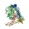
- PDB-9g4v: Group II intron assembly intermediate Domain 1 and 2 "Fully open"... -
+ Open data

- Basic information
| Entry | Database: PDB / ID: 9g4v | ||||||||||||||||||||||||||||||
|---|---|---|---|---|---|---|---|---|---|---|---|---|---|---|---|---|---|---|---|---|---|---|---|---|---|---|---|---|---|---|---|
| Title | Group II intron assembly intermediate Domain 1 and 2 "Fully open" state | ||||||||||||||||||||||||||||||
 Components Components | GROUP IIC INTRON | ||||||||||||||||||||||||||||||
 Keywords Keywords | RNA / RNA folding / protein-free RNA cryo-EM / Ribozyme / Metalloenzymes / Splicing | ||||||||||||||||||||||||||||||
| Function / homology | RNA / RNA (> 10) / RNA (> 100) Function and homology information Function and homology information | ||||||||||||||||||||||||||||||
| Biological species |  Oceanobacillus iheyensis (bacteria) Oceanobacillus iheyensis (bacteria) | ||||||||||||||||||||||||||||||
| Method | ELECTRON MICROSCOPY / single particle reconstruction / cryo EM / Resolution: 4.69 Å | ||||||||||||||||||||||||||||||
 Authors Authors | Jadhav, S.S. / Marcia, M. | ||||||||||||||||||||||||||||||
| Funding support |  France, 9items France, 9items
| ||||||||||||||||||||||||||||||
 Citation Citation | Journal: Nat Commun / Year: 2025 Title: Dynamic assembly of a large multidomain ribozyme visualized by cryo-electron microscopy. Authors: Shekhar Jadhav / Mauro Maiorca / Jacopo Manigrasso / Spandan Saha / Auriane Rakitch / Stefano Muscat / Thomas Mulvaney / Marco De Vivo / Maya Topf / Marco Marcia /    Abstract: Many RNAs rely on their 3D structures for function. While acquiring functional 3D structures, certain RNAs form misfolded, non-functional states ('kinetic traps'). Instead, other RNAs sequentially ...Many RNAs rely on their 3D structures for function. While acquiring functional 3D structures, certain RNAs form misfolded, non-functional states ('kinetic traps'). Instead, other RNAs sequentially assemble into their functional conformations over pre-folded scaffolds. Elucidating the principles of RNA sequential assembly is thus important to understand how RNAs avoid the formation of misfolded, non-functional states. Integrating single-particle electron cryomicroscopy (cryo-EM), image processing, in solution small-angle X-ray scattering (SAXS), EM-driven molecular dynamics (MD) simulations, structure-based mutagenesis, and enzymatic assays, we have visualized the sequential multidomain assembly of a self-splicing ribozyme of biomedical and bioengineering significance. Our work reveals a distinct dynamic interplay of helical subdomains in the ribozyme's 5'-terminal scaffold, which acts as a gate to control the docking of 3'-terminal domains. We identify specific conserved and functionally important secondary structure motifs as the key players for orchestrating the energetically inexpensive conformational changes that lead to the productive formation of the catalytic pocket. Our work provides a near-atomic resolution molecular movie of a large multidomain RNA assembling into its functionally active conformation and establishes a basis for understanding how RNA avoids the formation of non-functional 'kinetic traps'. | ||||||||||||||||||||||||||||||
| History |
|
- Structure visualization
| Structure viewer | Molecule: MolmilJmol/JSmol |
|---|
- Downloads & links
-Download
| PDBx/mmCIF format | 9g4v.cif.gz | 135.6 KB | Display | PDBx/mmCIF format |
|---|---|---|---|---|
| PDB format | pdb9g4v.ent.gz | 97.1 KB | Display | PDB format |
| PDBx/mmJSON format | 9g4v.json.gz | Tree view | PDBx/mmJSON format | |
| Others |  Other downloads Other downloads |
-Validation report
| Arichive directory | https://data.pdbj.org/pub/pdb/validation_reports/g4/9g4vftp://data.pdbj.org/pub/pdb/validation_reports/g4/9g4v | HTTPS FTP |
|---|
-Related structure data
| Related structure data |  51068MC  9g4iC  9g4jC  9g4lC  9g54C  9g56C M: map data used to model this data C: citing same article ( |
|---|---|
| Similar structure data |


-Links
 PDBj
PDBj


- Assembly
| Deposited unit | 
|
|---|---|
| 1 |
|
-Components
| #1: RNA chain | Mass: 96130.016 Da / Num. of mol.: 1 / Source method: obtained synthetically / Source: (synth.) Oceanobacillus iheyensis (bacteria) |
|---|---|
| Has protein modification | N |
-Experimental details
-Experiment
| Experiment | Method: ELECTRON MICROSCOPY |
|---|---|
| EM experiment | Aggregation state: PARTICLE / 3D reconstruction method: single particle reconstruction |
- Sample preparation
| Component | Name: GROUP IIC INTRON / Type: COMPLEX / Entity ID: all / Source: NATURAL |
|---|---|
| Molecular weight | Value: 0.101 MDa / Experimental value: NO |
| Source (natural) | Organism: Oceanobacillus iheyensis (bacteria) |
| Buffer solution | pH: 6.5 |
| Buffer component | Conc.: 0.5 uM Name: Sodium Chloride and 4-(2-sulfonatoethyl)morpholin-4-ium Formula: Na-MES |
| Specimen | Embedding applied: NO / Shadowing applied: NO / Staining applied: NO / Vitrification applied: YES |
| Specimen support | Grid material: COPPER / Grid mesh size: 300 divisions/in. / Grid type: Quantifoil R1.2/1.3 |
| Vitrification | Instrument: FEI VITROBOT MARK IV / Cryogen name: ETHANE / Humidity: 100 % |
- Electron microscopy imaging
| Experimental equipment |  Model: Titan Krios / Image courtesy: FEI Company |
|---|---|
| Microscopy | Model: FEI TITAN KRIOS |
| Electron gun | Electron source:  FIELD EMISSION GUN / Accelerating voltage: 300 kV / Illumination mode: FLOOD BEAM FIELD EMISSION GUN / Accelerating voltage: 300 kV / Illumination mode: FLOOD BEAM |
| Electron lens | Mode: BRIGHT FIELD / Nominal magnification: 105000 X / Nominal defocus max: 2200 nm / Nominal defocus min: 800 nm / Cs: 2.7 mm / Alignment procedure: COMA FREE |
| Specimen holder | Cryogen: NITROGEN / Specimen holder model: FEI TITAN KRIOS AUTOGRID HOLDER |
| Image recording | Electron dose: 38.07 e/Å2 / Detector mode: SUPER-RESOLUTION / Film or detector model: GATAN K3 (6k x 4k) |
| EM imaging optics | Energyfilter name: GIF Quantum LS / Energyfilter slit width: 20 eV |
- Processing
| EM software |
| ||||||||||||||||||||||||||||||||||||
|---|---|---|---|---|---|---|---|---|---|---|---|---|---|---|---|---|---|---|---|---|---|---|---|---|---|---|---|---|---|---|---|---|---|---|---|---|---|
| CTF correction | Type: PHASE FLIPPING AND AMPLITUDE CORRECTION | ||||||||||||||||||||||||||||||||||||
| Particle selection | Num. of particles selected: 306972 | ||||||||||||||||||||||||||||||||||||
| 3D reconstruction | Resolution: 4.69 Å / Resolution method: FSC 0.143 CUT-OFF / Num. of particles: 39075 / Symmetry type: POINT | ||||||||||||||||||||||||||||||||||||
| Atomic model building | Space: REAL | ||||||||||||||||||||||||||||||||||||
| Atomic model building | PDB-ID: 4FAQ Pdb chain-ID: A / Accession code: 4FAQ / Chain residue range: -4-289 / Pdb chain residue range: -4-289 / Source name: PDB / Type: experimental model |