 Movie
Movie Controller

[English] 日本語
 Yorodumi
- PDB-9g47: The structure of Candida albicans phosphoglucose isomerase in com... -
+ Open data

- Basic information
| Entry | Database: PDB / ID: 9g47 | |||||||||
|---|---|---|---|---|---|---|---|---|---|---|
| Title | The structure of Candida albicans phosphoglucose isomerase in complex with a fragment binder | |||||||||
 Components Components | Glucose-6-phosphate isomerase | |||||||||
 Keywords Keywords | ISOMERASE / PGI / Candida / fragment | |||||||||
| Function / homology |  Function and homology information Function and homology informationglucose-6-phosphate 1-epimerase activity / Isomerases; Racemases and epimerases; Acting on carbohydrates and derivatives / fungal biofilm matrix / racemase and epimerase activity, acting on carbohydrates and derivatives / glucose-6-phosphate isomerase / glucose-6-phosphate isomerase activity / glucose 6-phosphate metabolic process / carbohydrate derivative binding / monosaccharide binding / canonical glycolysis ...glucose-6-phosphate 1-epimerase activity / Isomerases; Racemases and epimerases; Acting on carbohydrates and derivatives / fungal biofilm matrix / racemase and epimerase activity, acting on carbohydrates and derivatives / glucose-6-phosphate isomerase / glucose-6-phosphate isomerase activity / glucose 6-phosphate metabolic process / carbohydrate derivative binding / monosaccharide binding / canonical glycolysis / glycolytic process / gluconeogenesis / mitochondrion / cytosol Similarity search - Function | |||||||||
| Biological species |  Candida albicans (yeast) Candida albicans (yeast) | |||||||||
| Method |  X-RAY DIFFRACTION / SYNCHROTRON / MOLECULAR REPLACEMENT / Resolution: 1.51 Å X-RAY DIFFRACTION / SYNCHROTRON / MOLECULAR REPLACEMENT / Resolution: 1.51 Å | |||||||||
 Authors Authors | Yan, K. | |||||||||
| Funding support |  United Kingdom, 2items United Kingdom, 2items
| |||||||||
 Citation Citation | Journal: Febs Lett. / Year: 2026 Title: Cell wall target fragment discovery using a low-cost, minimal fragment library. Authors: Yan, K. / Stanley, M. / Raimi, O. / Ferenbach, A.T. / Dorfmueller, H.C. / van Aalten, D.M.F. | |||||||||
| History |
|
- Structure visualization
| Structure viewer | Molecule: MolmilJmol/JSmol |
|---|
- Downloads & links
-Download
| PDBx/mmCIF format | 9g47.cif.gz | 252.4 KB | Display | PDBx/mmCIF format |
|---|---|---|---|---|
| PDB format | pdb9g47.ent.gz | Display | PDB format | |
| PDBx/mmJSON format | 9g47.json.gz | Tree view | PDBx/mmJSON format | |
| Others |  Other downloads Other downloads |
-Validation report
| Arichive directory | https://data.pdbj.org/pub/pdb/validation_reports/g4/9g47ftp://data.pdbj.org/pub/pdb/validation_reports/g4/9g47 | HTTPS FTP |
|---|
-Related structure data
| Related structure data |  9fztC  9g07C  9g45C  9g46C  9g4hC  9g4kC  9g4oC  9g53C  9g5aC  9g5fC  9g5oC  9g5yC  9g63C  9swyC C: citing same article ( |
|---|---|
| Similar structure data |
-Links
 PDBj
PDBj

- Assembly
| Deposited unit | 
| ||||||||
|---|---|---|---|---|---|---|---|---|---|
| 1 |
| ||||||||
| Unit cell |
|
-Components
| #1: Protein | Mass: 61670.438 Da / Num. of mol.: 2 Source method: isolated from a genetically manipulated source Source: (gene. exp.) Candida albicans (yeast) / Gene: PGI1, CAALFM_CR06340CA, CaO19.11369, CaO19.3888 / Production host:  #2: Sugar | 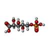  Type: saccharide / Mass: 246.109 Da / Num. of mol.: 2 / Source method: obtained synthetically / Formula: C5H11O9P Type: saccharide / Mass: 246.109 Da / Num. of mol.: 2 / Source method: obtained synthetically / Formula: C5H11O9P#3: Chemical | Mass: 146.146 Da / Num. of mol.: 2 / Source method: obtained synthetically / Formula: C8H6N2O / Feature type: SUBJECT OF INVESTIGATION #4: Water | ChemComp-HOH / |  Mass: 18.015 Da / Num. of mol.: 1085 / Source method: isolated from a natural source / Formula: H2O Mass: 18.015 Da / Num. of mol.: 1085 / Source method: isolated from a natural source / Formula: H2OHas ligand of interest | Y | Has protein modification | N | |
|---|
-Experimental details
-Experiment
| Experiment | Method: X-RAY DIFFRACTION / Number of used crystals: 1 |
|---|
- Sample preparation
| Crystal | Density Matthews: 2.41 Å3/Da / Density % sol: 48.94 % |
|---|---|
| Crystal grow | Temperature: 291 K / Method: vapor diffusion, sitting drop / Details: 0.1 M MgCl2, 0.1 M Hepes-NaOH pH 7.0, 21 % PEG4000 |
-Data collection
| Diffraction | Mean temperature: 100 K / Serial crystal experiment: N |
|---|---|
| Diffraction source | Source: SYNCHROTRON / Site: Diamond / Beamline: I04 / Wavelength: 0.95 Å |
| Detector | Type: DECTRIS EIGER2 XE 16M / Detector: PIXEL / Date: Jul 3, 2023 |
| Radiation | Protocol: SINGLE WAVELENGTH / Monochromatic (M) / Laue (L): M / Scattering type: x-ray |
| Radiation wavelength | Wavelength: 0.95 Å / Relative weight: 1 |
| Reflection | Resolution: 1.506→81.05 Å / Num. obs: 154412 / % possible obs: 84.2 % / Redundancy: 11.4 % / CC1/2: 1 / Net I/σ(I): 19.6 |
| Reflection shell | Resolution: 1.506→1.569 Å / Num. unique obs: 7721 / CC1/2: 0.983 |
- Processing
| Software |
| |||||||||||||||||||||||||||||||||||||||||||||||||||||||||||||||||||||||||||||||||||||||||||||||||||||||||||||||||||||||||||||||||||||||||||||||||||||||||||||||||||||||||||||||||||||||||||||||||||||||||||||||||||||||||
|---|---|---|---|---|---|---|---|---|---|---|---|---|---|---|---|---|---|---|---|---|---|---|---|---|---|---|---|---|---|---|---|---|---|---|---|---|---|---|---|---|---|---|---|---|---|---|---|---|---|---|---|---|---|---|---|---|---|---|---|---|---|---|---|---|---|---|---|---|---|---|---|---|---|---|---|---|---|---|---|---|---|---|---|---|---|---|---|---|---|---|---|---|---|---|---|---|---|---|---|---|---|---|---|---|---|---|---|---|---|---|---|---|---|---|---|---|---|---|---|---|---|---|---|---|---|---|---|---|---|---|---|---|---|---|---|---|---|---|---|---|---|---|---|---|---|---|---|---|---|---|---|---|---|---|---|---|---|---|---|---|---|---|---|---|---|---|---|---|---|---|---|---|---|---|---|---|---|---|---|---|---|---|---|---|---|---|---|---|---|---|---|---|---|---|---|---|---|---|---|---|---|---|---|---|---|---|---|---|---|---|---|---|---|---|---|---|---|---|
| Refinement | Method to determine structure: MOLECULAR REPLACEMENT / Resolution: 1.51→81.05 Å / SU ML: 0.11 / Cross valid method: FREE R-VALUE / σ(F): 1.34 / Phase error: 44.25 / Stereochemistry target values: ML
| |||||||||||||||||||||||||||||||||||||||||||||||||||||||||||||||||||||||||||||||||||||||||||||||||||||||||||||||||||||||||||||||||||||||||||||||||||||||||||||||||||||||||||||||||||||||||||||||||||||||||||||||||||||||||
| Solvent computation | Shrinkage radii: 0.9 Å / VDW probe radii: 1.11 Å / Solvent model: FLAT BULK SOLVENT MODEL | |||||||||||||||||||||||||||||||||||||||||||||||||||||||||||||||||||||||||||||||||||||||||||||||||||||||||||||||||||||||||||||||||||||||||||||||||||||||||||||||||||||||||||||||||||||||||||||||||||||||||||||||||||||||||
| Refinement step | Cycle: LAST / Resolution: 1.51→81.05 Å
| |||||||||||||||||||||||||||||||||||||||||||||||||||||||||||||||||||||||||||||||||||||||||||||||||||||||||||||||||||||||||||||||||||||||||||||||||||||||||||||||||||||||||||||||||||||||||||||||||||||||||||||||||||||||||
| Refine LS restraints |
| |||||||||||||||||||||||||||||||||||||||||||||||||||||||||||||||||||||||||||||||||||||||||||||||||||||||||||||||||||||||||||||||||||||||||||||||||||||||||||||||||||||||||||||||||||||||||||||||||||||||||||||||||||||||||
| LS refinement shell |
|
