 Movie
Movie Controller

[English] 日本語
 Yorodumi
- PDB-9g3b: Crystal Structure of the artificial protein METP in complex with ... -
+ Open data

- Basic information
| Entry | Database: PDB / ID: 9g3b | ||||||
|---|---|---|---|---|---|---|---|
| Title | Crystal Structure of the artificial protein METP in complex with cadmium ion at different temperature, 130 K | ||||||
 Components Components | METP ARTIFICIAL PROTEIN | ||||||
 Keywords Keywords | DE NOVO PROTEIN / de novo design / room temperature / cadmium-sulphur cluster / denovo protein | ||||||
| Function / homology | :  Function and homology information Function and homology information | ||||||
| Biological species | synthetic construct (others) | ||||||
| Method |  X-RAY DIFFRACTION / SYNCHROTRON / SAD / Resolution: 1.9 Å X-RAY DIFFRACTION / SYNCHROTRON / SAD / Resolution: 1.9 Å | ||||||
 Authors Authors | Di Costanzo, L. / La Gatta, S. / Chino, M. | ||||||
| Funding support |  Italy, 1items Italy, 1items
| ||||||
 Citation Citation | Journal: J.Inorg.Biochem. / Year: 2025 Title: Structural insights into temperature-dependent dynamics of METPsc1, a miniaturized electron-transfer protein. Authors: Di Costanzo, L.F. / Sgueglia, G. / Orlando, C. / Polentarutti, M. / Leone, L. / La Gatta, S. / De Fenza, M. / De Gioia, L. / Lombardi, A. / Arrigoni, F. / Chino, M. | ||||||
| History |
|
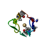
- Structure visualization
| Structure viewer | Molecule: MolmilJmol/JSmol |
|---|
- Downloads & links
-Download
| PDBx/mmCIF format | 9g3b.cif.gz | 25.7 KB | Display | PDBx/mmCIF format |
|---|---|---|---|---|
| PDB format | pdb9g3b.ent.gz | 12.9 KB | Display | PDB format |
| PDBx/mmJSON format | 9g3b.json.gz | Tree view | PDBx/mmJSON format | |
| Others |  Other downloads Other downloads |
-Validation report
| Arichive directory | https://data.pdbj.org/pub/pdb/validation_reports/g3/9g3bftp://data.pdbj.org/pub/pdb/validation_reports/g3/9g3b | HTTPS FTP |
|---|
-Related structure data


-Links
 PDBj
PDBj
- Assembly
| Deposited unit | 
| ||||||||||||
|---|---|---|---|---|---|---|---|---|---|---|---|---|---|
| 1 |
| ||||||||||||
| Unit cell |
| ||||||||||||
| Components on special symmetry positions |
|
-Components
| #1: Protein/peptide | Mass: 2955.254 Da / Num. of mol.: 1 / Source method: obtained synthetically / Source: (synth.) synthetic construct (others) |
|---|---|
| #2: Chemical | ChemComp-CD /   Mass: 112.411 Da / Num. of mol.: 1 / Source method: obtained synthetically / Formula: Cd / Feature type: SUBJECT OF INVESTIGATION Mass: 112.411 Da / Num. of mol.: 1 / Source method: obtained synthetically / Formula: Cd / Feature type: SUBJECT OF INVESTIGATION |
| #3: Water | ChemComp-HOH /  Mass: 18.015 Da / Num. of mol.: 18 / Source method: isolated from a natural source / Formula: H2O Mass: 18.015 Da / Num. of mol.: 18 / Source method: isolated from a natural source / Formula: H2O |
| Has ligand of interest | Y |
| Has protein modification | Y |
-Experimental details
-Experiment
| Experiment | Method: X-RAY DIFFRACTION / Number of used crystals: 1 |
|---|
- Sample preparation
| Crystal | Density Matthews: 2.8 Å3/Da / Density % sol: 56.6 % |
|---|---|
| Crystal grow | Temperature: 298 K / Method: vapor diffusion, hanging drop / pH: 7.5 Details: drop containing 2.0 μL of 1:1 (v/v) mixture of protein solution (10 mg/mL, 7 mM DTT, 4 mM CdCl2) and 2.0 μL of precipitant buffer (0.1 M HEPES at pH 7.5, 1.4 M sodium citrate ...Details: drop containing 2.0 μL of 1:1 (v/v) mixture of protein solution (10 mg/mL, 7 mM DTT, 4 mM CdCl2) and 2.0 μL of precipitant buffer (0.1 M HEPES at pH 7.5, 1.4 M sodium citrate tribasic dihydrate) was equilibrated against 0.5 mL reservoir of precipitant buffer |
-Data collection
| Diffraction | Mean temperature: 100 K / Serial crystal experiment: N |
|---|---|
| Diffraction source | Source: SYNCHROTRON / Site: ELETTRA / Beamline: 5.2R / Wavelength: 0.534 Å |
| Detector | Type: DECTRIS PILATUS3 6M / Detector: PIXEL / Date: Jan 1, 2000 |
| Radiation | Protocol: SINGLE WAVELENGTH / Monochromatic (M) / Laue (L): M / Scattering type: x-ray |
| Radiation wavelength | Wavelength: 0.534 Å / Relative weight: 1 |
| Reflection | Resolution: 1.7→29.37 Å / Num. obs: 8740 / % possible obs: 95.1 % / Redundancy: 2.2 % / Biso Wilson estimate: 15.15 Å2 / CC1/2: 0.995 / Rmerge(I) obs: 0.079 / Net I/σ(I): 8.82 |
| Reflection shell | Resolution: 1.7→1.8 Å / Num. unique obs: 450 / CC1/2: 0.903 / % possible all: 96.3 |
- Processing
| Software |
| ||||||||||||||||||||||||
|---|---|---|---|---|---|---|---|---|---|---|---|---|---|---|---|---|---|---|---|---|---|---|---|---|---|
| Refinement | Method to determine structure: SAD / Resolution: 1.9→29.37 Å / SU ML: 0.2588 / Cross valid method: FREE R-VALUE / σ(F): 1.34 / Phase error: 9.8902 Stereochemistry target values: GeoStd + Monomer Library + CDL v1.2
| ||||||||||||||||||||||||
| Solvent computation | Shrinkage radii: 0.9 Å / VDW probe radii: 1.1 Å / Solvent model: FLAT BULK SOLVENT MODEL | ||||||||||||||||||||||||
| Displacement parameters | Biso mean: 20.31 Å2 | ||||||||||||||||||||||||
| Refinement step | Cycle: LAST / Resolution: 1.9→29.37 Å
| ||||||||||||||||||||||||
| Refine LS restraints |
| ||||||||||||||||||||||||
| LS refinement shell | Resolution: 1.9→29.37 Å
|
