- PDB-9g13: VHH H3-2 in complex with Tau C-terminal peptide -
+
Open data
ID or keywords:
Loading...
-
Basic information
Entry
Database: PDB / ID: 9g13
Title
VHH H3-2 in complex with Tau C-terminal peptide
Components
Isoform Tau-F of Microtubule-associated protein tau
VHH H3-2
Keywords
IMMUNE SYSTEM / VHH / Nanobody / Tau
Function / homology
Function and homology information
plus-end-directed organelle transport along microtubule / histone-dependent DNA binding / neurofibrillary tangle assembly / positive regulation of diacylglycerol kinase activity / axonal transport / negative regulation of establishment of protein localization to mitochondrion / neurofibrillary tangle / positive regulation of protein localization to synapse / microtubule lateral binding / tubulin complex ...plus-end-directed organelle transport along microtubule / histone-dependent DNA binding / neurofibrillary tangle assembly / positive regulation of diacylglycerol kinase activity / axonal transport / negative regulation of establishment of protein localization to mitochondrion / neurofibrillary tangle / positive regulation of protein localization to synapse / microtubule lateral binding / tubulin complex / phosphatidylinositol bisphosphate binding / main axon / regulation of long-term synaptic depression / negative regulation of kinase activity / negative regulation of tubulin deacetylation / generation of neurons / rRNA metabolic process / internal protein amino acid acetylation / regulation of chromosome organization / regulation of mitochondrial fission / axonal transport of mitochondrion / axon development / intracellular distribution of mitochondria / central nervous system neuron development / regulation of microtubule polymerization / microtubule polymerization / lipoprotein particle binding / minor groove of adenine-thymine-rich DNA binding / dynactin binding / glial cell projection / negative regulation of mitochondrial membrane potential / apolipoprotein binding / protein polymerization / axolemma / negative regulation of mitochondrial fission / Caspase-mediated cleavage of cytoskeletal proteins / regulation of microtubule polymerization or depolymerization / positive regulation of axon extension / regulation of microtubule cytoskeleton organization / Activation of AMPK downstream of NMDARs / positive regulation of protein localization / regulation of cellular response to heat / cytoplasmic microtubule organization / stress granule assembly / supramolecular fiber organization / regulation of calcium-mediated signaling / axon cytoplasm / somatodendritic compartment / positive regulation of microtubule polymerization / cellular response to brain-derived neurotrophic factor stimulus / synapse assembly / phosphatidylinositol binding / nuclear periphery / cellular response to nerve growth factor stimulus / positive regulation of superoxide anion generation / protein phosphatase 2A binding / regulation of autophagy / astrocyte activation / response to lead ion / microglial cell activation / synapse organization / Hsp90 protein binding / protein homooligomerization / PKR-mediated signaling / regulation of synaptic plasticity / memory / activation of cysteine-type endopeptidase activity involved in apoptotic process / microtubule cytoskeleton organization / SH3 domain binding / cellular response to reactive oxygen species / cytoplasmic ribonucleoprotein granule / microtubule cytoskeleton / neuron projection development / cell-cell signaling / protein-folding chaperone binding / protein-macromolecule adaptor activity / single-stranded DNA binding / actin binding / cellular response to heat / cell body / growth cone / microtubule binding / double-stranded DNA binding / microtubule / sequence-specific DNA binding / amyloid fibril formation / dendritic spine / learning or memory / nuclear speck / neuron projection / membrane raft / axon / negative regulation of gene expression / neuronal cell body / dendrite / DNA damage response / protein kinase binding / enzyme binding / mitochondrion / DNA binding Similarity search - Function
: / Microtubule associated protein, tubulin-binding repeat / Microtubule-associated protein Tau / Tau and MAP protein, tubulin-binding repeat / Tau and MAP proteins tubulin-binding repeat signature. / Tau and MAP proteins tubulin-binding repeat profile. Similarity search - Domain/homology
A: VHH H3-2 B: Isoform Tau-F of Microtubule-associated protein tau E: VHH H3-2 F: Isoform Tau-F of Microtubule-associated protein tau C: VHH H3-2 D: Isoform Tau-F of Microtubule-associated protein tau G: VHH H3-2 H: Isoform Tau-F of Microtubule-associated protein tau
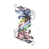
A: VHH H3-2 B: Isoform Tau-F of Microtubule-associated protein tau C: VHH H3-2 D: Isoform Tau-F of Microtubule-associated protein tau
defined by author&software
Evidence: gel filtration
31.8 kDa, 4 polymers
Theoretical mass
Number of molelcules
Total (without water)
31,839
4
Polymers
31,839
4
Non-polymers
0
0
Water
72
4
Type
Name
Symmetry operation
Number
identity operation
1_555
x,y,z
1
Buried area
8540 Å2
ΔGint
-49 kcal/mol
Surface area
14020 Å2
Method
PISA
2
E: VHH H3-2 F: Isoform Tau-F of Microtubule-associated protein tau G: VHH H3-2 H: Isoform Tau-F of Microtubule-associated protein tau
defined by author&software
Evidence: gel filtration
31.8 kDa, 4 polymers
Theoretical mass
Number of molelcules
Total (without water)
31,839
4
Polymers
31,839
4
Non-polymers
0
0
Water
72
4
Type
Name
Symmetry operation
Number
identity operation
1_555
x,y,z
1
Buried area
8390 Å2
ΔGint
-46 kcal/mol
Surface area
13790 Å2
Method
PISA
Unit cell
Length a, b, c (Å)
70.394, 76.346, 95.565
Angle α, β, γ (deg.)
90.000, 90.000, 90.000
Int Tables number
19
Space group name H-M
P212121
-
Components
#1: Antibody
VHHH3-2
Mass: 14270.698 Da / Num. of mol.: 4 Source method: isolated from a genetically manipulated source Source: (gene. exp.) Lama glama (llama) / Production host: Escherichia coli (E. coli)
In the structure databanks used in Yorodumi, some data are registered as the other names, "COVID-19 virus" and "2019-nCoV". Here are the details of the virus and the list of structure data.
Jan 31, 2019. EMDB accession codes are about to change! (news from PDBe EMDB page)
EMDB accession codes are about to change! (news from PDBe EMDB page)
The allocation of 4 digits for EMDB accession codes will soon come to an end. Whilst these codes will remain in use, new EMDB accession codes will include an additional digit and will expand incrementally as the available range of codes is exhausted. The current 4-digit format prefixed with “EMD-” (i.e. EMD-XXXX) will advance to a 5-digit format (i.e. EMD-XXXXX), and so on. It is currently estimated that the 4-digit codes will be depleted around Spring 2019, at which point the 5-digit format will come into force.
The EM Navigator/Yorodumi systems omit the EMD- prefix.
Related info.:Q: What is EMD? / ID/Accession-code notation in Yorodumi/EM Navigator
Yorodumi is a browser for structure data from EMDB, PDB, SASBDB, etc.
This page is also the successor to EM Navigator detail page, and also detail information page/front-end page for Omokage search.
The word "yorodu" (or yorozu) is an old Japanese word meaning "ten thousand". "mi" (miru) is to see.
Related info.:EMDB / PDB / SASBDB / Comparison of 3 databanks / Yorodumi Search / Aug 31, 2016. New EM Navigator & Yorodumi / Yorodumi Papers / Jmol/JSmol / Function and homology information / Changes in new EM Navigator and Yorodumi
 Movie
Movie Controller


 Open data
 Basic information
 Components
 Keywords
 Function and homology information
 Homo sapiens (human)
 X-RAY DIFFRACTION /
  Authors
 France, 2items
  Citation
 Structure visualization
 Downloads & links
 Other downloads
 PDBj
PDBj


 Assembly
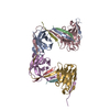


 Mass: 18.015 Da / Num. of mol.: 470 / Source method: isolated from a natural source / Formula: H2O
 Sample preparation
 Processing