 Movie
Movie Controller


+ Open data

- Basic information
| Entry | Database: PDB / ID: 9fym | ||||||
|---|---|---|---|---|---|---|---|
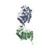
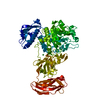
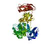
| Title | Lacto-N-biosidase from Treponema denticola ATCC 35405 | ||||||
 Components Components | Glycoside hydrolase family 20 catalytic domain-containing protein | ||||||
 Keywords Keywords | HYDROLASE / lacto-N-biosidase / GH20 | ||||||
| Function / homology |  Function and homology information Function and homology informationN-acetyllactosaminidase / lacto-N-biosidase / beta-N-acetylhexosaminidase activity / carbohydrate metabolic process / metal ion binding Similarity search - Function | ||||||
| Biological species |  Treponema denticola ATCC 35405 (bacteria) Treponema denticola ATCC 35405 (bacteria) | ||||||
| Method |  X-RAY DIFFRACTION / SYNCHROTRON / MOLECULAR REPLACEMENT / Resolution: 1.34 Å X-RAY DIFFRACTION / SYNCHROTRON / MOLECULAR REPLACEMENT / Resolution: 1.34 Å | ||||||
 Authors Authors | Vuillemin, M. / Siebenhaar, S. / Zeuner, B. / Morth, J.P. | ||||||
| Funding support |  Denmark, 1items Denmark, 1items
| ||||||
 Citation Citation | Journal: Chembiochem / Year: 2025 Title: Discovery of Lacto-N-Biosidases and a Novel N-Acetyllactosaminidase Activity in the CAZy Family GH20: Functional Diversity and Structural Insights. Authors: Vuillemin, M. / Muschiol, J. / Zhang, Y. / Holck, J. / Barrett, K. / Preben Morth, J. / Meyer, A.S. / Zeuner, B. | ||||||
| History |
|
- Structure visualization
| Structure viewer | Molecule: MolmilJmol/JSmol |
|---|
- Downloads & links
-Download
| PDBx/mmCIF format | 9fym.cif.gz | 181 KB | Display | PDBx/mmCIF format |
|---|---|---|---|---|
| PDB format | pdb9fym.ent.gz | 116.2 KB | Display | PDB format |
| PDBx/mmJSON format | 9fym.json.gz | Tree view | PDBx/mmJSON format | |
| Others |  Other downloads Other downloads |
-Validation report
| Arichive directory | https://data.pdbj.org/pub/pdb/validation_reports/fy/9fymftp://data.pdbj.org/pub/pdb/validation_reports/fy/9fym | HTTPS FTP |
|---|
-Related structure data
-Links
 PDBj
PDBj


- Assembly
| Deposited unit | 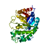
| ||||||||||||
|---|---|---|---|---|---|---|---|---|---|---|---|---|---|
| 1 |
| ||||||||||||
| Unit cell |
|
-Components
| #1: Protein | Mass: 37816.203 Da / Num. of mol.: 1 Source method: isolated from a genetically manipulated source Details: Signal peptide has been removed, additional GS (from Thrombin cleavage). Source: (gene. exp.) Treponema denticola ATCC 35405 (bacteria)Gene: TDE_1723 / Production host: | ||||||||
|---|---|---|---|---|---|---|---|---|---|
| #2: Chemical | ChemComp-IMD /   Mass: 69.085 Da / Num. of mol.: 1 / Source method: obtained synthetically / Formula: C3H5N2 Mass: 69.085 Da / Num. of mol.: 1 / Source method: obtained synthetically / Formula: C3H5N2 | ||||||||
| #3: Chemical |   Mass: 22.990 Da / Num. of mol.: 2 / Source method: obtained synthetically / Formula: Na Mass: 22.990 Da / Num. of mol.: 2 / Source method: obtained synthetically / Formula: Na#4: Chemical |   Mass: 65.409 Da / Num. of mol.: 2 / Source method: isolated from a natural source / Formula: Zn Mass: 65.409 Da / Num. of mol.: 2 / Source method: isolated from a natural source / Formula: Zn#5: Water | ChemComp-HOH / |  Mass: 18.015 Da / Num. of mol.: 352 / Source method: isolated from a natural source / Formula: H2O Mass: 18.015 Da / Num. of mol.: 352 / Source method: isolated from a natural source / Formula: H2OHas ligand of interest | N | Has protein modification | N | |
-Experimental details
-Experiment
| Experiment | Method: X-RAY DIFFRACTION / Number of used crystals: 1 |
|---|
- Sample preparation
| Crystal | Density Matthews: 2.11 Å3/Da / Density % sol: 41.76 % |
|---|---|
| Crystal grow | Temperature: 294.15 K / Method: vapor diffusion, sitting drop / pH: 7.5 Details: 10% w/v PEG 8000, 9% ethylene glycol, 0.1M HEPES pH 7.5 |
-Data collection
| Diffraction | Mean temperature: 100 K / Serial crystal experiment: N |
|---|---|
| Diffraction source | Source: SYNCHROTRON / Site: ESRF  / Beamline: ID30B / Wavelength: 1.0332 Å / Beamline: ID30B / Wavelength: 1.0332 Å |
| Detector | Type: DECTRIS PILATUS 6M / Detector: PIXEL / Date: Feb 1, 2023 |
| Radiation | Protocol: SINGLE WAVELENGTH / Monochromatic (M) / Laue (L): M / Scattering type: x-ray |
| Radiation wavelength | Wavelength: 1.0332 Å / Relative weight: 1 |
| Reflection | Resolution: 1.34→48.15 Å / Num. obs: 77556 / % possible obs: 99.58 % / Redundancy: 8.2 % / Biso Wilson estimate: 17.71 Å2 / CC1/2: 0.998 / CC star: 1 / Rmerge(I) obs: 0.08324 / Rpim(I) all: 0.03112 / Rrim(I) all: 0.08909 / Net I/σ(I): 11.31 |
| Reflection shell | Resolution: 1.34→1.39 Å / Redundancy: 7.1 % / Rmerge(I) obs: 1 / Mean I/σ(I) obs: 0.83 / Num. unique obs: 7469 / CC1/2: 0.464 / Rpim(I) all: 1 / Rrim(I) all: 1 / % possible all: 97.97 |
- Processing
| Software |
| |||||||||||||||||||||||||||||||||||||||||||||||||||||||||||||||||||||||||||||||||||||||||||||||||||||||||||||||||||||||||||||||||||||||||||||||||||||||||||||||||||||||||||||||||||||||||||||||||||||||||||||||||||||||||
|---|---|---|---|---|---|---|---|---|---|---|---|---|---|---|---|---|---|---|---|---|---|---|---|---|---|---|---|---|---|---|---|---|---|---|---|---|---|---|---|---|---|---|---|---|---|---|---|---|---|---|---|---|---|---|---|---|---|---|---|---|---|---|---|---|---|---|---|---|---|---|---|---|---|---|---|---|---|---|---|---|---|---|---|---|---|---|---|---|---|---|---|---|---|---|---|---|---|---|---|---|---|---|---|---|---|---|---|---|---|---|---|---|---|---|---|---|---|---|---|---|---|---|---|---|---|---|---|---|---|---|---|---|---|---|---|---|---|---|---|---|---|---|---|---|---|---|---|---|---|---|---|---|---|---|---|---|---|---|---|---|---|---|---|---|---|---|---|---|---|---|---|---|---|---|---|---|---|---|---|---|---|---|---|---|---|---|---|---|---|---|---|---|---|---|---|---|---|---|---|---|---|---|---|---|---|---|---|---|---|---|---|---|---|---|---|---|---|---|
| Refinement | Method to determine structure: MOLECULAR REPLACEMENT / Resolution: 1.34→48.15 Å / SU ML: 0.175 / Cross valid method: FREE R-VALUE / σ(F): 1.33 / Phase error: 23.4484 Stereochemistry target values: GeoStd + Monomer Library + CDL v1.2
| |||||||||||||||||||||||||||||||||||||||||||||||||||||||||||||||||||||||||||||||||||||||||||||||||||||||||||||||||||||||||||||||||||||||||||||||||||||||||||||||||||||||||||||||||||||||||||||||||||||||||||||||||||||||||
| Solvent computation | Shrinkage radii: 0.9 Å / VDW probe radii: 1.1 Å / Solvent model: FLAT BULK SOLVENT MODEL | |||||||||||||||||||||||||||||||||||||||||||||||||||||||||||||||||||||||||||||||||||||||||||||||||||||||||||||||||||||||||||||||||||||||||||||||||||||||||||||||||||||||||||||||||||||||||||||||||||||||||||||||||||||||||
| Displacement parameters | Biso mean: 26.93 Å2 | |||||||||||||||||||||||||||||||||||||||||||||||||||||||||||||||||||||||||||||||||||||||||||||||||||||||||||||||||||||||||||||||||||||||||||||||||||||||||||||||||||||||||||||||||||||||||||||||||||||||||||||||||||||||||
| Refinement step | Cycle: LAST / Resolution: 1.34→48.15 Å
| |||||||||||||||||||||||||||||||||||||||||||||||||||||||||||||||||||||||||||||||||||||||||||||||||||||||||||||||||||||||||||||||||||||||||||||||||||||||||||||||||||||||||||||||||||||||||||||||||||||||||||||||||||||||||
| Refine LS restraints |
| |||||||||||||||||||||||||||||||||||||||||||||||||||||||||||||||||||||||||||||||||||||||||||||||||||||||||||||||||||||||||||||||||||||||||||||||||||||||||||||||||||||||||||||||||||||||||||||||||||||||||||||||||||||||||
| LS refinement shell |
|
