 Movie
Movie Controller

[English] 日本語
 Yorodumi
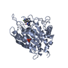
- PDB-9fxh: Crystal structure of cobalt(II)-substituted double mutant Y115E Y... -
+ Open data

- Basic information
| Entry | Database: PDB / ID: 9fxh | ||||||
|---|---|---|---|---|---|---|---|
| Title | Crystal structure of cobalt(II)-substituted double mutant Y115E Y117E human Glutaminyl Cyclase | ||||||
 Components Components | Glutaminyl-peptide cyclotransferase | ||||||
 Keywords Keywords | TRANSFERASE / human glutaminyl cyclase / acyltransferase / hQC / cobalt | ||||||
| Function / homology |  Function and homology information Function and homology informationpeptidyl-pyroglutamic acid biosynthetic process, using glutaminyl-peptide cyclotransferase / glutaminyl-peptide cyclotransferase / glutaminyl-peptide cyclotransferase activity / protein modification process / specific granule lumen / tertiary granule lumen / ficolin-1-rich granule lumen / Neutrophil degranulation / extracellular exosome / extracellular region / zinc ion binding Similarity search - Function | ||||||
| Biological species |  Homo sapiens (human) Homo sapiens (human) | ||||||
| Method |  X-RAY DIFFRACTION / SYNCHROTRON / MOLECULAR REPLACEMENT / Resolution: 2.3 Å X-RAY DIFFRACTION / SYNCHROTRON / MOLECULAR REPLACEMENT / Resolution: 2.3 Å | ||||||
 Authors Authors | Tassone, G. / Pozzi, C. / Mangani, S. | ||||||
| Funding support |  Italy, 1items Italy, 1items
| ||||||
 Citation Citation | Journal: Int J Mol Sci / Year: 2024 Title: Metal Ion Binding to Human Glutaminyl Cyclase: A Structural Perspective. Authors: Tassone, G. / Pozzi, C. / Mangani, S. | ||||||
| History |
|
- Structure visualization
| Structure viewer | Molecule: MolmilJmol/JSmol |
|---|
- Downloads & links
-Download
| PDBx/mmCIF format | 9fxh.cif.gz | 229.9 KB | Display | PDBx/mmCIF format |
|---|---|---|---|---|
| PDB format | pdb9fxh.ent.gz | 181.5 KB | Display | PDB format |
| PDBx/mmJSON format | 9fxh.json.gz | Tree view | PDBx/mmJSON format | |
| Others |  Other downloads Other downloads |
-Validation report
| Arichive directory | https://data.pdbj.org/pub/pdb/validation_reports/fx/9fxhftp://data.pdbj.org/pub/pdb/validation_reports/fx/9fxh | HTTPS FTP |
|---|
-Related structure data


-Links
 PDBj
PDBj

- Assembly
| Deposited unit | 
| |||||||||||||||
|---|---|---|---|---|---|---|---|---|---|---|---|---|---|---|---|---|
| 1 | 
| |||||||||||||||
| 2 | 
| |||||||||||||||
| 3 | 
| |||||||||||||||
| Unit cell |
| |||||||||||||||
| Components on special symmetry positions |
|
-Components
| #1: Protein | Mass: 38894.789 Da / Num. of mol.: 3 / Mutation: Y115E, Y117E Source method: isolated from a genetically manipulated source Source: (gene. exp.) Homo sapiens (human) / Gene: QPCT / Plasmid: PLASMID / Details (production host): pQE-80L / Production host:  References: UniProt: Q16769, glutaminyl-peptide cyclotransferase #2: Chemical |   Mass: 58.933 Da / Num. of mol.: 3 / Source method: obtained synthetically / Formula: Co / Feature type: SUBJECT OF INVESTIGATION Mass: 58.933 Da / Num. of mol.: 3 / Source method: obtained synthetically / Formula: Co / Feature type: SUBJECT OF INVESTIGATION#3: Chemical |   Mass: 96.063 Da / Num. of mol.: 3 / Source method: obtained synthetically / Formula: SO4 Mass: 96.063 Da / Num. of mol.: 3 / Source method: obtained synthetically / Formula: SO4#4: Chemical | ChemComp-GOL / |   Mass: 92.094 Da / Num. of mol.: 1 / Source method: isolated from a natural source / Formula: C3H8O3 Mass: 92.094 Da / Num. of mol.: 1 / Source method: isolated from a natural source / Formula: C3H8O3#5: Water | ChemComp-HOH / |  Mass: 18.015 Da / Num. of mol.: 1070 / Source method: isolated from a natural source / Formula: H2O Mass: 18.015 Da / Num. of mol.: 1070 / Source method: isolated from a natural source / Formula: H2OHas ligand of interest | Y | |
|---|
-Experimental details
-Experiment
| Experiment | Method: X-RAY DIFFRACTION / Number of used crystals: 1 |
|---|
- Sample preparation
| Crystal | Density Matthews: 2.63 Å3/Da / Density % sol: 53.18 % |
|---|---|
| Crystal grow | Temperature: 281 K / Method: vapor diffusion, sitting drop / pH: 6 / Details: 0.1-0.2 M ammonium sulphate and 0.1 M MES, pH 6.0 |
-Data collection
| Diffraction | Mean temperature: 100 K / Serial crystal experiment: N |
|---|---|
| Diffraction source | Source: SYNCHROTRON / Site: Diamond  / Beamline: I04 / Wavelength: 0.95374 Å / Beamline: I04 / Wavelength: 0.95374 Å |
| Detector | Type: DECTRIS EIGER2 XE 16M / Detector: PIXEL / Date: Dec 3, 2023 |
| Radiation | Monochromator: Si(111) / Protocol: MAD / Monochromatic (M) / Laue (L): M / Scattering type: x-ray |
| Radiation wavelength | Wavelength: 0.95374 Å / Relative weight: 1 |
| Reflection | Resolution: 2.3→61.76 Å / Num. obs: 50664 / % possible obs: 95.2 % / Observed criterion σ(I): 2 / Redundancy: 5.4 % / Biso Wilson estimate: 19.9 Å2 / CC1/2: 0.989 / Rmerge(I) obs: 0.157 / Rpim(I) all: 0.112 / Rrim(I) all: 0.194 / Net I/σ(I): 7 |
| Reflection shell | Resolution: 2.3→2.38 Å / Rmerge(I) obs: 0.621 / Mean I/σ(I) obs: 2.2 / Num. unique obs: 4761 / CC1/2: 0.747 / Rpim(I) all: 0.452 / Rrim(I) all: 0.772 / % possible all: 97 |
- Processing
| Software |
| ||||||||||||||||||||||||||||||||||||||||||||||||||||||||||||||||||||||||||||||||||||||||||||||||||||||||||||||||||||||||||||||||||||||||||||||||||||||||||||||||||||||||||||||||||||||
|---|---|---|---|---|---|---|---|---|---|---|---|---|---|---|---|---|---|---|---|---|---|---|---|---|---|---|---|---|---|---|---|---|---|---|---|---|---|---|---|---|---|---|---|---|---|---|---|---|---|---|---|---|---|---|---|---|---|---|---|---|---|---|---|---|---|---|---|---|---|---|---|---|---|---|---|---|---|---|---|---|---|---|---|---|---|---|---|---|---|---|---|---|---|---|---|---|---|---|---|---|---|---|---|---|---|---|---|---|---|---|---|---|---|---|---|---|---|---|---|---|---|---|---|---|---|---|---|---|---|---|---|---|---|---|---|---|---|---|---|---|---|---|---|---|---|---|---|---|---|---|---|---|---|---|---|---|---|---|---|---|---|---|---|---|---|---|---|---|---|---|---|---|---|---|---|---|---|---|---|---|---|---|---|
| Refinement | Method to determine structure: MOLECULAR REPLACEMENT / Resolution: 2.3→61.76 Å / Cor.coef. Fo:Fc: 0.936 / Cor.coef. Fo:Fc free: 0.902 / SU B: 9.825 / SU ML: 0.222 / Cross valid method: THROUGHOUT / ESU R: 0.406 / ESU R Free: 0.262 / Stereochemistry target values: MAXIMUM LIKELIHOOD
| ||||||||||||||||||||||||||||||||||||||||||||||||||||||||||||||||||||||||||||||||||||||||||||||||||||||||||||||||||||||||||||||||||||||||||||||||||||||||||||||||||||||||||||||||||||||
| Solvent computation | Ion probe radii: 0.8 Å / Shrinkage radii: 0.8 Å / VDW probe radii: 1.2 Å / Solvent model: MASK | ||||||||||||||||||||||||||||||||||||||||||||||||||||||||||||||||||||||||||||||||||||||||||||||||||||||||||||||||||||||||||||||||||||||||||||||||||||||||||||||||||||||||||||||||||||||
| Displacement parameters | Biso mean: 29.135 Å2
| ||||||||||||||||||||||||||||||||||||||||||||||||||||||||||||||||||||||||||||||||||||||||||||||||||||||||||||||||||||||||||||||||||||||||||||||||||||||||||||||||||||||||||||||||||||||
| Refine analyze | Luzzati coordinate error obs: 0.29 Å | ||||||||||||||||||||||||||||||||||||||||||||||||||||||||||||||||||||||||||||||||||||||||||||||||||||||||||||||||||||||||||||||||||||||||||||||||||||||||||||||||||||||||||||||||||||||
| Refinement step | Cycle: 1 / Resolution: 2.3→61.76 Å
| ||||||||||||||||||||||||||||||||||||||||||||||||||||||||||||||||||||||||||||||||||||||||||||||||||||||||||||||||||||||||||||||||||||||||||||||||||||||||||||||||||||||||||||||||||||||
| Refine LS restraints |
|