 Movie
Movie Controller


+ Open data

- Basic information
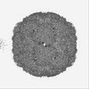
| Entry | Database: PDB / ID: 9fx1 | ||||||||||||||||||
|---|---|---|---|---|---|---|---|---|---|---|---|---|---|---|---|---|---|---|---|
| Title | CryoEM structure of RV-A89 | ||||||||||||||||||
 Components Components | (Capsid protein ...) x 4 | ||||||||||||||||||
 Keywords Keywords | VIRUS / RV-A89 / rhinovirus | ||||||||||||||||||
| Function / homology |  Function and homology information Function and homology informationsymbiont-mediated suppression of host cytoplasmic pattern recognition receptor signaling pathway via inhibition of RIG-I activity / picornain 2A / symbiont-mediated suppression of host mRNA export from nucleus / symbiont genome entry into host cell via pore formation in plasma membrane / picornain 3C / T=pseudo3 icosahedral viral capsid / host cell cytoplasmic vesicle membrane / ribonucleoside triphosphate phosphatase activity / nucleoside-triphosphate phosphatase / channel activity ...symbiont-mediated suppression of host cytoplasmic pattern recognition receptor signaling pathway via inhibition of RIG-I activity / picornain 2A / symbiont-mediated suppression of host mRNA export from nucleus / symbiont genome entry into host cell via pore formation in plasma membrane / picornain 3C / T=pseudo3 icosahedral viral capsid / host cell cytoplasmic vesicle membrane / ribonucleoside triphosphate phosphatase activity / nucleoside-triphosphate phosphatase / channel activity / monoatomic ion transmembrane transport / DNA replication / RNA helicase activity / endocytosis involved in viral entry into host cell / symbiont-mediated activation of host autophagy / RNA-directed RNA polymerase / cysteine-type endopeptidase activity / viral RNA genome replication / RNA-directed RNA polymerase activity / virion attachment to host cell / DNA-templated transcription / host cell nucleus / structural molecule activity / proteolysis / RNA binding / zinc ion binding / ATP binding Similarity search - Function | ||||||||||||||||||
| Biological species |  Human rhinovirus 89 ATCC VR-1199 Human rhinovirus 89 ATCC VR-1199 | ||||||||||||||||||
| Method | ELECTRON MICROSCOPY / single particle reconstruction / cryo EM / Resolution: 1.76 Å | ||||||||||||||||||
 Authors Authors | Wald, J. / Goessweiner-Mohr, N. / Blaas, D. / Marlovits, T.C. | ||||||||||||||||||
| Funding support |  Germany, 5items Germany, 5items
| ||||||||||||||||||
 Citation Citation | Journal: Sci Rep / Year: 2024 Title: DMSO might impact ligand binding, capsid stability, and RNA interaction in viral preparations. Authors: Jiri Wald / Nikolaus Goessweiner-Mohr / Antonio Real-Hohn / Dieter Blaas / Thomas C Marlovits /  Abstract: Dimethyl sulfoxide (DMSO) is a widely used solvent in drug research. However, recent studies indicate that even at low concentration DMSO might cause structural changes of proteins and RNA. The ...Dimethyl sulfoxide (DMSO) is a widely used solvent in drug research. However, recent studies indicate that even at low concentration DMSO might cause structural changes of proteins and RNA. The pyrazolopyrimidine antiviral OBR-5-340 dissolved in DMSO inhibits rhinovirus-B5 infection yet is inactive against RV-A89. This is consistent with our structural observation that OBR-5-340 is only visible at the pocket factor site in rhinovirus-B5 and not in RV-A89, where the hydrophobic pocket is collapsed. Here, we analyze the impact of DMSO in RV-A89 by high-resolution cryo-electron microscopy. Our 1.76 Å cryo-EM reconstruction of RV-A89 in plain buffer, without DMSO, reveals that the pocket-factor binding site is occupied by myristate and that the previously observed local heterogeneity at protein-RNA interfaces is absent. These findings suggest that DMSO elutes the pocket factor, leading to a collapse of the hydrophobic pocket of RV-A89. Consequently, the conformational heterogeneity observed at the RNA-protein interface in the presence of DMSO likely results from increased capsid flexibility due to the absence of the pocket factor and DMSO-induced affinity modifications. This local asymmetry may promote a directional release of the RNA genome during infection. | ||||||||||||||||||
| History |
|
- Structure visualization
| Structure viewer | Molecule: MolmilJmol/JSmol |
|---|
- Downloads & links
-Download
| PDBx/mmCIF format | 9fx1.cif.gz | 298.4 KB | Display | PDBx/mmCIF format |
|---|---|---|---|---|
| PDB format | pdb9fx1.ent.gz | 244.5 KB | Display | PDB format |
| PDBx/mmJSON format | 9fx1.json.gz | Tree view | PDBx/mmJSON format | |
| Others |  Other downloads Other downloads |
-Validation report
| Arichive directory | https://data.pdbj.org/pub/pdb/validation_reports/fx/9fx1ftp://data.pdbj.org/pub/pdb/validation_reports/fx/9fx1 | HTTPS FTP |
|---|
-Related structure data
| Related structure data |  50840MC  9fx9C M: map data used to model this data C: citing same article ( |
|---|---|
| Similar structure data |
-Links
 PDBj
PDBj


- Assembly
| Deposited unit | 
|
|---|---|
| 1 | x 60
|
-Components
-Capsid protein ... , 4 types, 4 molecules ABCD
| #1: Protein | Mass: 31920.547 Da / Num. of mol.: 1 Source method: isolated from a genetically manipulated source Source: (gene. exp.) Human rhinovirus 89 ATCC VR-1199 / Production host: Human rhinovirus 89 ATCC VR-1199 / References: UniProt: P07210 |
|---|---|
| #2: Protein | Mass: 28097.393 Da / Num. of mol.: 1 Source method: isolated from a genetically manipulated source Source: (gene. exp.) Human rhinovirus 89 ATCC VR-1199 / Production host: Human rhinovirus 89 ATCC VR-1199 / References: UniProt: P07210 |
| #3: Protein | Mass: 23824.178 Da / Num. of mol.: 1 Source method: isolated from a genetically manipulated source Source: (gene. exp.) Human rhinovirus 89 ATCC VR-1199 / Production host: Human rhinovirus 89 ATCC VR-1199 / References: UniProt: P07210 |
| #4: Protein/peptide | Mass: 1830.952 Da / Num. of mol.: 1 Source method: isolated from a genetically manipulated source Source: (gene. exp.) Human rhinovirus 89 ATCC VR-1199 / Production host: Human rhinovirus 89 ATCC VR-1199 / References: UniProt: P07210 |
-Non-polymers , 2 types, 205 molecules 

| #5: Chemical | ChemComp-DAO /  Mass: 200.318 Da / Num. of mol.: 1 / Source method: obtained synthetically / Formula: C12H24O2 / Feature type: SUBJECT OF INVESTIGATION Mass: 200.318 Da / Num. of mol.: 1 / Source method: obtained synthetically / Formula: C12H24O2 / Feature type: SUBJECT OF INVESTIGATION |
|---|---|
| #6: Water | ChemComp-HOH / Mass: 18.015 Da / Num. of mol.: 204 / Source method: isolated from a natural source / Formula: H2O |
-Details
| Has ligand of interest | Y |
|---|---|
| Has protein modification | N |
-Experimental details
-Experiment
| Experiment | Method: ELECTRON MICROSCOPY |
|---|---|
| EM experiment | Aggregation state: PARTICLE / 3D reconstruction method: single particle reconstruction |
- Sample preparation
| Component | Name: Human rhinovirus 89 ATCC VR-1199 / Type: VIRUS / Entity ID: #1-#4 / Source: NATURAL |
|---|---|
| Molecular weight | Experimental value: NO |
| Source (natural) | Organism: Human rhinovirus 89 ATCC VR-1199 |
| Details of virus | Empty: NO / Enveloped: NO / Isolate: SEROTYPE / Type: VIRION |
| Natural host | Organism: Homo sapiens |
| Buffer solution | pH: 7.4 |
| Specimen | Embedding applied: NO / Shadowing applied: NO / Staining applied: NO / Vitrification applied: YES |
| Specimen support | Grid material: COPPER / Grid mesh size: 300 divisions/in. / Grid type: Quantifoil R2/1 |
| Vitrification | Instrument: FEI VITROBOT MARK IV / Cryogen name: ETHANE-PROPANE / Humidity: 100 % / Chamber temperature: 277 K |
- Electron microscopy imaging
| Experimental equipment |  Model: Titan Krios / Image courtesy: FEI Company |
|---|---|
| Microscopy | Model: TFS KRIOS |
| Electron gun | Electron source:  FIELD EMISSION GUN / Accelerating voltage: 300 kV / Illumination mode: FLOOD BEAM FIELD EMISSION GUN / Accelerating voltage: 300 kV / Illumination mode: FLOOD BEAM |
| Electron lens | Mode: BRIGHT FIELD / Nominal magnification: 105000 X / Nominal defocus max: 1500 nm / Nominal defocus min: 500 nm / Cs: 2.7 mm / C2 aperture diameter: 70 µm / Alignment procedure: ZEMLIN TABLEAU |
| Specimen holder | Cryogen: NITROGEN / Specimen holder model: FEI TITAN KRIOS AUTOGRID HOLDER |
| Image recording | Average exposure time: 2 sec. / Electron dose: 44 e/Å2 / Film or detector model: GATAN K3 BIOQUANTUM (6k x 4k) / Num. of grids imaged: 1 / Num. of real images: 4860 |
- Processing
| EM software |
| ||||||||||||||||||||||||||||||||||||||||||||
|---|---|---|---|---|---|---|---|---|---|---|---|---|---|---|---|---|---|---|---|---|---|---|---|---|---|---|---|---|---|---|---|---|---|---|---|---|---|---|---|---|---|---|---|---|---|
| CTF correction | Type: NONE | ||||||||||||||||||||||||||||||||||||||||||||
| Symmetry | Point symmetry: I (icosahedral) | ||||||||||||||||||||||||||||||||||||||||||||
| 3D reconstruction | Resolution: 1.76 Å / Resolution method: FSC 0.143 CUT-OFF / Num. of particles: 97123 / Symmetry type: POINT | ||||||||||||||||||||||||||||||||||||||||||||
| Atomic model building | Protocol: OTHER |
