 Movie
Movie Controller

[English] 日本語
 Yorodumi
- PDB-9fsa: Cell wall anchoring domain of the surface layer protein of Methan... -
+ Open data

- Basic information
| Entry | Database: PDB / ID: 9fsa | ||||||
|---|---|---|---|---|---|---|---|
| Title | Cell wall anchoring domain of the surface layer protein of Methanococcus voltae (aa 24-75; 484-576) | ||||||
 Components Components | S-layer protein | ||||||
 Keywords Keywords | STRUCTURAL PROTEIN / Surface layer protein / self assembly / cell wall binding / anchoring / symmetry | ||||||
| Function / homology | S-layer protein / S-layer protein, N-terminal / S-layer protein, C-terminal / S-layer like family, central region / S-layer like family, outer domain / S-layer / cell wall organization / extracellular region / S-layer protein Function and homology information Function and homology information | ||||||
| Biological species |  Methanococcus voltae (archaea) Methanococcus voltae (archaea) | ||||||
| Method |  X-RAY DIFFRACTION / MOLECULAR REPLACEMENT / Resolution: 2.05 Å X-RAY DIFFRACTION / MOLECULAR REPLACEMENT / Resolution: 2.05 Å | ||||||
 Authors Authors | Grininger, C. / Sagmeister, T. / Pavkov-Keller, T. | ||||||
| Funding support |  Austria, 1items Austria, 1items
| ||||||
 Citation Citation | Journal: Nat Commun / Year: 2024 Title: SymProFold: Structural prediction of symmetrical biological assemblies. Authors: Buhlheller, C. / Sagmeister, T. / Grininger, C. / Gubensak, N. / Sleytr, U.B. / Uson, I. / Pavkov-Keller, T. | ||||||
| History |
|
- Structure visualization
| Structure viewer | Molecule: MolmilJmol/JSmol |
|---|
- Downloads & links
-Download
| PDBx/mmCIF format | 9fsa.cif.gz | 59.9 KB | Display | PDBx/mmCIF format |
|---|---|---|---|---|
| PDB format | pdb9fsa.ent.gz | 41.3 KB | Display | PDB format |
| PDBx/mmJSON format | 9fsa.json.gz | Tree view | PDBx/mmJSON format | |
| Others |  Other downloads Other downloads |
-Validation report
| Arichive directory | https://data.pdbj.org/pub/pdb/validation_reports/fs/9fsaftp://data.pdbj.org/pub/pdb/validation_reports/fs/9fsa | HTTPS FTP |
|---|
-Related structure data
| Similar structure data |
|---|
-Links
 PDBj
PDBj- Assembly
| Deposited unit | 
| ||||||||
|---|---|---|---|---|---|---|---|---|---|
| 1 | 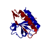
| ||||||||
| Unit cell |
|
-Components
| #1: Protein | Mass: 15757.842 Da / Num. of mol.: 1 Source method: isolated from a genetically manipulated source Source: (gene. exp.) Methanococcus voltae (archaea) / Gene: slaProduction host:  References: UniProt: Q50833 |
|---|---|
| #2: Water | ChemComp-HOH /  Mass: 18.015 Da / Num. of mol.: 14 / Source method: isolated from a natural source / Formula: H2O Mass: 18.015 Da / Num. of mol.: 14 / Source method: isolated from a natural source / Formula: H2O |
-Experimental details
-Experiment
| Experiment | Method: X-RAY DIFFRACTION / Number of used crystals: 1 |
|---|
- Sample preparation
| Crystal | Density Matthews: 2.18 Å3/Da / Density % sol: 43.54 % |
|---|---|
| Crystal grow | Temperature: 293 K / Method: vapor diffusion, sitting drop / pH: 4 Details: JCSG+ eco condition C2 1.0 M LiCl, 0.1 M citrate pH 4.0, 20 % PEG 6000 0.3 ul condition + 0.3 ul protein solution |
-Data collection
| Diffraction | Mean temperature: 100 K / Serial crystal experiment: N |
|---|---|
| Diffraction source | Source: LIQUID ANODE / Type: Excillum MetalJet D2+ 70 kV / Wavelength: 1.3414 Å |
| Detector | Type: Bruker PHOTON III / Detector: PIXEL / Date: Jun 12, 2024 |
| Radiation | Protocol: SINGLE WAVELENGTH / Monochromatic (M) / Laue (L): M / Scattering type: x-ray |
| Radiation wavelength | Wavelength: 1.3414 Å / Relative weight: 1 |
| Reflection | Resolution: 2.05→46.34 Å / Num. obs: 4977 / % possible obs: 90.7 % / Redundancy: 23.5 % / CC1/2: 0.996 / Rmerge(I) obs: 0.332 / Net I/σ(I): 8.2 |
| Reflection shell | Resolution: 2.05→2.24 Å / Redundancy: 15.8 % / Rmerge(I) obs: 1.8 / Mean I/σ(I) obs: 1.6 / Num. unique obs: 291 / CC1/2: 0.65 / % possible all: 70.1 |
- Processing
| Software |
| |||||||||||||||||||||||||||||||||||||||||||||||||||||||||||||||||||||||||||||||||||||||||||||||||||||||||||||||||||||||||||||||||||||||||||||||||||||||||||||||||||||||||||||||||||||||||||||||||||||||||||||||||||||||||||||||||||||||
|---|---|---|---|---|---|---|---|---|---|---|---|---|---|---|---|---|---|---|---|---|---|---|---|---|---|---|---|---|---|---|---|---|---|---|---|---|---|---|---|---|---|---|---|---|---|---|---|---|---|---|---|---|---|---|---|---|---|---|---|---|---|---|---|---|---|---|---|---|---|---|---|---|---|---|---|---|---|---|---|---|---|---|---|---|---|---|---|---|---|---|---|---|---|---|---|---|---|---|---|---|---|---|---|---|---|---|---|---|---|---|---|---|---|---|---|---|---|---|---|---|---|---|---|---|---|---|---|---|---|---|---|---|---|---|---|---|---|---|---|---|---|---|---|---|---|---|---|---|---|---|---|---|---|---|---|---|---|---|---|---|---|---|---|---|---|---|---|---|---|---|---|---|---|---|---|---|---|---|---|---|---|---|---|---|---|---|---|---|---|---|---|---|---|---|---|---|---|---|---|---|---|---|---|---|---|---|---|---|---|---|---|---|---|---|---|---|---|---|---|---|---|---|---|---|---|---|---|---|---|---|---|---|
| Refinement | Method to determine structure: MOLECULAR REPLACEMENT / Resolution: 2.05→46.34 Å / Cor.coef. Fo:Fc: 0.882 / Cor.coef. Fo:Fc free: 0.873 / SU B: 6.53 / SU ML: 0.179 / Cross valid method: FREE R-VALUE / ESU R: 0.686 / ESU R Free: 0.324 Details: Hydrogens have been added in their riding positions
| |||||||||||||||||||||||||||||||||||||||||||||||||||||||||||||||||||||||||||||||||||||||||||||||||||||||||||||||||||||||||||||||||||||||||||||||||||||||||||||||||||||||||||||||||||||||||||||||||||||||||||||||||||||||||||||||||||||||
| Solvent computation | Ion probe radii: 0.8 Å / Shrinkage radii: 0.8 Å / VDW probe radii: 1.2 Å / Solvent model: MASK BULK SOLVENT | |||||||||||||||||||||||||||||||||||||||||||||||||||||||||||||||||||||||||||||||||||||||||||||||||||||||||||||||||||||||||||||||||||||||||||||||||||||||||||||||||||||||||||||||||||||||||||||||||||||||||||||||||||||||||||||||||||||||
| Displacement parameters | Biso mean: 22.898 Å2
| |||||||||||||||||||||||||||||||||||||||||||||||||||||||||||||||||||||||||||||||||||||||||||||||||||||||||||||||||||||||||||||||||||||||||||||||||||||||||||||||||||||||||||||||||||||||||||||||||||||||||||||||||||||||||||||||||||||||
| Refinement step | Cycle: LAST / Resolution: 2.05→46.34 Å
| |||||||||||||||||||||||||||||||||||||||||||||||||||||||||||||||||||||||||||||||||||||||||||||||||||||||||||||||||||||||||||||||||||||||||||||||||||||||||||||||||||||||||||||||||||||||||||||||||||||||||||||||||||||||||||||||||||||||
| Refine LS restraints |
| |||||||||||||||||||||||||||||||||||||||||||||||||||||||||||||||||||||||||||||||||||||||||||||||||||||||||||||||||||||||||||||||||||||||||||||||||||||||||||||||||||||||||||||||||||||||||||||||||||||||||||||||||||||||||||||||||||||||
| LS refinement shell | Refine-ID: X-RAY DIFFRACTION / Total num. of bins used: 20
|
