 Movie
Movie Controller


+ Open data

- Basic information
| Entry | Database: PDB / ID: 9fro | ||||||
|---|---|---|---|---|---|---|---|
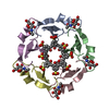
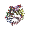
| Title | Crystal structure of Pent - p-sulfonatocalix[6]arene complex | ||||||
 Components Components | Beta propeller | ||||||
 Keywords Keywords | SUGAR BINDING PROTEIN / Complex / Beta-propeller / Lectin / Pentamer | ||||||
| Function / homology | Tachylectin 2 / Tachylectin 2 superfamily / Tachylectin / p-sulfonatocalix[6]arene / 2-acetamido-2-deoxy-alpha-D-glucopyranose / Beta propeller Function and homology information Function and homology information | ||||||
| Biological species |  Enterobacteria phage L1 (virus) Enterobacteria phage L1 (virus) | ||||||
| Method |  X-RAY DIFFRACTION / SYNCHROTRON / MOLECULAR REPLACEMENT / Resolution: 1.71 Å X-RAY DIFFRACTION / SYNCHROTRON / MOLECULAR REPLACEMENT / Resolution: 1.71 Å | ||||||
 Authors Authors | Flood, R.J. / Crowley, P.B. | ||||||
| Funding support |  Ireland, 1items Ireland, 1items
| ||||||
 Citation Citation | Journal: ACS Macro Lett / Year: 2024 Title: A Macrocycle-Mediated Protein Cage. Authors: Flood, R.J. / Thureau, A. / Crowley, P.B. #1: Journal: Biomacromolecules / Year: 2024Title: Multivalent Calixarene Complexation of a Designed Pentameric Lectin. Authors: Flood, R.J. / Cerofolini, L. / Fragai, M. / Crowley, P.B. | ||||||
| History |
|
- Structure visualization
| Structure viewer | Molecule: MolmilJmol/JSmol |
|---|
- Downloads & links
-Download
| PDBx/mmCIF format | 9fro.cif.gz | 84.7 KB | Display | PDBx/mmCIF format |
|---|---|---|---|---|
| PDB format | pdb9fro.ent.gz | 53.7 KB | Display | PDB format |
| PDBx/mmJSON format | 9fro.json.gz | Tree view | PDBx/mmJSON format | |
| Others |  Other downloads Other downloads |
-Validation report
| Arichive directory | https://data.pdbj.org/pub/pdb/validation_reports/fr/9froftp://data.pdbj.org/pub/pdb/validation_reports/fr/9fro | HTTPS FTP |
|---|
-Related structure data
| Related structure data | |
|---|---|
| Similar structure data |

-Links
 PDBj
PDBj


- Assembly
| Deposited unit | 
| ||||||||||||
|---|---|---|---|---|---|---|---|---|---|---|---|---|---|
| 1 |
| ||||||||||||
| Unit cell |
| ||||||||||||
| Components on special symmetry positions |
|
-Components
| #1: Protein/peptide | Mass: 5327.980 Da / Num. of mol.: 5 Source method: isolated from a genetically manipulated source Source: (gene. exp.) Enterobacteria phage L1 (virus) / Production host:  #2: Sugar | ChemComp-NDG /   Type: D-saccharide, alpha linking / Mass: 221.208 Da / Num. of mol.: 4 / Source method: obtained synthetically / Formula: C8H15NO6 Type: D-saccharide, alpha linking / Mass: 221.208 Da / Num. of mol.: 4 / Source method: obtained synthetically / Formula: C8H15NO6#3: Chemical | ChemComp-FWQ /   Mass: 1111.063 Da / Num. of mol.: 4 / Source method: obtained synthetically / Formula: C42H30O24S6 / Feature type: SUBJECT OF INVESTIGATION Mass: 1111.063 Da / Num. of mol.: 4 / Source method: obtained synthetically / Formula: C42H30O24S6 / Feature type: SUBJECT OF INVESTIGATION#4: Water | ChemComp-HOH / |  Mass: 18.015 Da / Num. of mol.: 135 / Source method: isolated from a natural source / Formula: H2O Mass: 18.015 Da / Num. of mol.: 135 / Source method: isolated from a natural source / Formula: H2OHas ligand of interest | Y | Has protein modification | N | |
|---|
-Experimental details
-Experiment
| Experiment | Method: X-RAY DIFFRACTION / Number of used crystals: 1 |
|---|
- Sample preparation
| Crystal | Density Matthews: 2.78 Å3/Da / Density % sol: 55 % / Description: bipyramidal |
|---|---|
| Crystal grow | Temperature: 293 K / Method: vapor diffusion, hanging drop / pH: 7 Details: 1.2 M ammonium sulfate 0.1 M sodium malonate; pH 7.0 |
-Data collection
| Diffraction | Mean temperature: 100 K / Serial crystal experiment: N |
|---|---|
| Diffraction source | Source: SYNCHROTRON / Site: SOLEIL  / Beamline: PROXIMA 2 / Wavelength: 0.98011 Å / Beamline: PROXIMA 2 / Wavelength: 0.98011 Å |
| Detector | Type: DECTRIS EIGER X 9M / Detector: PIXEL / Date: Mar 25, 2023 |
| Radiation | Protocol: SINGLE WAVELENGTH / Monochromatic (M) / Laue (L): M / Scattering type: x-ray |
| Radiation wavelength | Wavelength: 0.98011 Å / Relative weight: 1 |
| Reflection | Resolution: 1.71→56.19 Å / Num. obs: 31640 / % possible obs: 100 % / Redundancy: 12.2 % / Biso Wilson estimate: 21.84 Å2 / CC1/2: 0.998 / Rmerge(I) obs: 0.123 / Rpim(I) all: 0.037 / Rrim(I) all: 0.129 / Net I/σ(I): 12.4 |
| Reflection shell | Resolution: 1.712→1.742 Å / Rmerge(I) obs: 1.438 / Mean I/σ(I) obs: 2.1 / Num. unique obs: 1593 / CC1/2: 0.779 / Rpim(I) all: 0.439 / Rrim(I) all: 1.505 / % possible all: 100 |
- Processing
| Software |
| ||||||||||||||||||||||||||||||||||||||||||||||||||||||||||||||||||||||||||||||||||||
|---|---|---|---|---|---|---|---|---|---|---|---|---|---|---|---|---|---|---|---|---|---|---|---|---|---|---|---|---|---|---|---|---|---|---|---|---|---|---|---|---|---|---|---|---|---|---|---|---|---|---|---|---|---|---|---|---|---|---|---|---|---|---|---|---|---|---|---|---|---|---|---|---|---|---|---|---|---|---|---|---|---|---|---|---|---|
| Refinement | Method to determine structure: MOLECULAR REPLACEMENT / Resolution: 1.71→56.19 Å / SU ML: 0.2117 / Cross valid method: FREE R-VALUE / σ(F): 1.36 / Phase error: 23.0286 Stereochemistry target values: GeoStd + Monomer Library + CDL v1.2
| ||||||||||||||||||||||||||||||||||||||||||||||||||||||||||||||||||||||||||||||||||||
| Solvent computation | Shrinkage radii: 0.9 Å / VDW probe radii: 1.1 Å / Solvent model: FLAT BULK SOLVENT MODEL | ||||||||||||||||||||||||||||||||||||||||||||||||||||||||||||||||||||||||||||||||||||
| Displacement parameters | Biso mean: 27.78 Å2 | ||||||||||||||||||||||||||||||||||||||||||||||||||||||||||||||||||||||||||||||||||||
| Refinement step | Cycle: LAST / Resolution: 1.71→56.19 Å
| ||||||||||||||||||||||||||||||||||||||||||||||||||||||||||||||||||||||||||||||||||||
| Refine LS restraints |
| ||||||||||||||||||||||||||||||||||||||||||||||||||||||||||||||||||||||||||||||||||||
| LS refinement shell |
|
