 Movie
Movie Controller


+ Open data

- Basic information
| Entry | Database: PDB / ID: 9frn | ||||||
|---|---|---|---|---|---|---|---|
| Title | The RSL-D46H - sulfonato-calix[8]arene complex, acetate pH 4.0 | ||||||
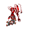
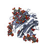
 Components Components | Fucose-binding lectin protein | ||||||
 Keywords Keywords | SUGAR BINDING PROTEIN / Assembly / Beta-propeller / Lectin / Trimer | ||||||
| Function / homology | Fucose-specific lectin / Fungal fucose-specific lectin / carbohydrate binding / metal ion binding / beta-D-fructopyranose / sulfonato-calix[8]arene / Fucose-binding lectin protein Function and homology information Function and homology information | ||||||
| Biological species |  Ralstonia solanacearum (bacteria) Ralstonia solanacearum (bacteria) | ||||||
| Method |  X-RAY DIFFRACTION / SYNCHROTRON / MOLECULAR REPLACEMENT / Resolution: 1.87 Å X-RAY DIFFRACTION / SYNCHROTRON / MOLECULAR REPLACEMENT / Resolution: 1.87 Å | ||||||
 Authors Authors | Flood, R.J. / Crowley, P.B. | ||||||
| Funding support |  Ireland, 1items Ireland, 1items
| ||||||
 Citation Citation | Journal: Cryst Growth Des / Year: 2024 Title: Supramolecular Synthons in Protein-Ligand Frameworks. Authors: Flood, R.J. / Mockler, N.M. / Thureau, A. / Malinska, M. / Crowley, P.B. | ||||||
| History |
|
- Structure visualization
| Structure viewer | Molecule: MolmilJmol/JSmol |
|---|
- Downloads & links
-Download
| PDBx/mmCIF format | 9frn.cif.gz | 165.3 KB | Display | PDBx/mmCIF format |
|---|---|---|---|---|
| PDB format | pdb9frn.ent.gz | 108.6 KB | Display | PDB format |
| PDBx/mmJSON format | 9frn.json.gz | Tree view | PDBx/mmJSON format | |
| Others |  Other downloads Other downloads |
-Validation report
| Arichive directory | https://data.pdbj.org/pub/pdb/validation_reports/fr/9frnftp://data.pdbj.org/pub/pdb/validation_reports/fr/9frn | HTTPS FTP |
|---|
-Related structure data


-Links
 PDBj
PDBj- Assembly
| Deposited unit | 
| ||||||||||||
|---|---|---|---|---|---|---|---|---|---|---|---|---|---|
| 1 | 
| ||||||||||||
| 2 | 
| ||||||||||||
| Unit cell |
|
-Components
| #1: Protein | Mass: 9756.621 Da / Num. of mol.: 6 Source method: isolated from a genetically manipulated source Details: D46H mutant of RSL / Source: (gene. exp.) Ralstonia solanacearum (bacteria) / Gene: E7Z57_08365, HF909_06975, LBW55_09125, RUN39_v1_50103 / Production host: #2: Chemical | ChemComp-EVB / 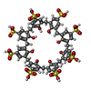  Mass: 1489.481 Da / Num. of mol.: 4 / Source method: obtained synthetically / Formula: C56H48O32S8 / Feature type: SUBJECT OF INVESTIGATION Mass: 1489.481 Da / Num. of mol.: 4 / Source method: obtained synthetically / Formula: C56H48O32S8 / Feature type: SUBJECT OF INVESTIGATION#3: Sugar | ChemComp-BDF /   Type: D-saccharide, beta linking / Mass: 180.156 Da / Num. of mol.: 12 / Source method: obtained synthetically / Formula: C6H12O6 Type: D-saccharide, beta linking / Mass: 180.156 Da / Num. of mol.: 12 / Source method: obtained synthetically / Formula: C6H12O6#4: Water | ChemComp-HOH / |  Mass: 18.015 Da / Num. of mol.: 406 / Source method: isolated from a natural source / Formula: H2O Mass: 18.015 Da / Num. of mol.: 406 / Source method: isolated from a natural source / Formula: H2OHas ligand of interest | Y | Has protein modification | N | |
|---|
-Experimental details
-Experiment
| Experiment | Method: X-RAY DIFFRACTION / Number of used crystals: 1 |
|---|
- Sample preparation
| Crystal | Density Matthews: 2.66 Å3/Da / Density % sol: 54 % |
|---|---|
| Crystal grow | Temperature: 277.15 K / Method: batch mode / pH: 4 / Details: 20 mM sodium acetate pH 4.0 50 mM sodium chloride |
-Data collection
| Diffraction | Mean temperature: 100 K / Serial crystal experiment: N |
|---|---|
| Diffraction source | Source: SYNCHROTRON / Site: SOLEIL  / Beamline: PROXIMA 2 / Wavelength: 0.98011 Å / Beamline: PROXIMA 2 / Wavelength: 0.98011 Å |
| Detector | Type: DECTRIS EIGER X 9M / Detector: PIXEL / Date: Nov 16, 2021 |
| Radiation | Protocol: SINGLE WAVELENGTH / Monochromatic (M) / Laue (L): M / Scattering type: x-ray |
| Radiation wavelength | Wavelength: 0.98011 Å / Relative weight: 1 |
| Reflection | Resolution: 1.87→47.46 Å / Num. obs: 48037 / % possible obs: 97 % / Redundancy: 3.4 % / Biso Wilson estimate: 22.94 Å2 / CC1/2: 0.995 / Rmerge(I) obs: 0.073 / Rpim(I) all: 0.046 / Rrim(I) all: 0.086 / Net I/σ(I): 5.6 |
| Reflection shell | Resolution: 1.87→1.9 Å / Redundancy: 3.6 % / Rmerge(I) obs: 0.416 / Mean I/σ(I) obs: 2.1 / Num. unique obs: 2445 / CC1/2: 0.895 / Rpim(I) all: 0.256 / Rrim(I) all: 0.489 / % possible all: 96.4 |
- Processing
| Software |
| |||||||||||||||||||||||||||||||||||||||||||||||||||||||||||||||||||||||||||||||||||||||||||||||||||||||||||||||||||||||
|---|---|---|---|---|---|---|---|---|---|---|---|---|---|---|---|---|---|---|---|---|---|---|---|---|---|---|---|---|---|---|---|---|---|---|---|---|---|---|---|---|---|---|---|---|---|---|---|---|---|---|---|---|---|---|---|---|---|---|---|---|---|---|---|---|---|---|---|---|---|---|---|---|---|---|---|---|---|---|---|---|---|---|---|---|---|---|---|---|---|---|---|---|---|---|---|---|---|---|---|---|---|---|---|---|---|---|---|---|---|---|---|---|---|---|---|---|---|---|---|---|
| Refinement | Method to determine structure: MOLECULAR REPLACEMENT / Resolution: 1.87→47.45 Å / SU ML: 0.2638 / Cross valid method: FREE R-VALUE / σ(F): 1.97 / Phase error: 28.5891 Stereochemistry target values: GeoStd + Monomer Library + CDL v1.2
| |||||||||||||||||||||||||||||||||||||||||||||||||||||||||||||||||||||||||||||||||||||||||||||||||||||||||||||||||||||||
| Solvent computation | Shrinkage radii: 0.9 Å / VDW probe radii: 1.1 Å / Solvent model: FLAT BULK SOLVENT MODEL | |||||||||||||||||||||||||||||||||||||||||||||||||||||||||||||||||||||||||||||||||||||||||||||||||||||||||||||||||||||||
| Displacement parameters | Biso mean: 29.64 Å2 | |||||||||||||||||||||||||||||||||||||||||||||||||||||||||||||||||||||||||||||||||||||||||||||||||||||||||||||||||||||||
| Refinement step | Cycle: LAST / Resolution: 1.87→47.45 Å
| |||||||||||||||||||||||||||||||||||||||||||||||||||||||||||||||||||||||||||||||||||||||||||||||||||||||||||||||||||||||
| Refine LS restraints |
| |||||||||||||||||||||||||||||||||||||||||||||||||||||||||||||||||||||||||||||||||||||||||||||||||||||||||||||||||||||||
| LS refinement shell |
|
