 Movie
Movie Controller


+ Open data

- Basic information
| Entry | Database: PDB / ID: 9fr5 | ||||||
|---|---|---|---|---|---|---|---|
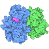
| Title | Crystal structure of human GSK3B in complex with ARN25697 | ||||||
 Components Components | Glycogen synthase kinase-3 beta | ||||||
 Keywords Keywords | TRANSFERASE / kinase inhibitors / multitarget compounds / drug discovery / central nervous system / tauopathies | ||||||
| Function / homology |  Function and homology information Function and homology informationnegative regulation of glycogen (starch) synthase activity / neuron projection organization / regulation of microtubule anchoring at centrosome / negative regulation of mesenchymal stem cell differentiation / negative regulation of type B pancreatic cell development / regulation of protein export from nucleus / superior temporal gyrus development / positive regulation of protein localization to cilium / negative regulation of glycogen biosynthetic process / negative regulation of TORC2 signaling ...negative regulation of glycogen (starch) synthase activity / neuron projection organization / regulation of microtubule anchoring at centrosome / negative regulation of mesenchymal stem cell differentiation / negative regulation of type B pancreatic cell development / regulation of protein export from nucleus / superior temporal gyrus development / positive regulation of protein localization to cilium / negative regulation of glycogen biosynthetic process / negative regulation of TORC2 signaling / beta-arrestin-dependent dopamine receptor signaling pathway / negative regulation of dopaminergic neuron differentiation / positive regulation of protein localization to centrosome / maintenance of cell polarity / positive regulation of cilium assembly / heart valve development / CRMPs in Sema3A signaling / tau-protein kinase / beta-catenin destruction complex / APC truncation mutants have impaired AXIN binding / AXIN missense mutants destabilize the destruction complex / Truncations of AMER1 destabilize the destruction complex / positive regulation of mitochondrial outer membrane permeabilization involved in apoptotic signaling pathway / negative regulation of calcineurin-NFAT signaling cascade / Maturation of nucleoprotein / cellular response to interleukin-3 / Beta-catenin phosphorylation cascade / Signaling by GSK3beta mutants / CTNNB1 S33 mutants aren't phosphorylated / CTNNB1 S37 mutants aren't phosphorylated / CTNNB1 S45 mutants aren't phosphorylated / CTNNB1 T41 mutants aren't phosphorylated / negative regulation of TOR signaling / regulation of long-term synaptic potentiation / Wnt signalosome / regulation of microtubule-based process / AKT phosphorylates targets in the cytosol / Disassembly of the destruction complex and recruitment of AXIN to the membrane / regulation of axon extension / positive regulation of protein binding / regulation of neuron projection development / negative regulation of protein localization to nucleus / Maturation of nucleoprotein / glycogen metabolic process / ER overload response / negative regulation of epithelial to mesenchymal transition / positive regulation of cell-matrix adhesion / tau-protein kinase activity / regulation of axonogenesis / establishment of cell polarity / regulation of dendrite morphogenesis / Constitutive Signaling by AKT1 E17K in Cancer / protein kinase A catalytic subunit binding / canonical Wnt signaling pathway / dynactin binding / epithelial to mesenchymal transition / Regulation of HSF1-mediated heat shock response / negative regulation of extrinsic apoptotic signaling pathway via death domain receptors / extrinsic apoptotic signaling pathway in absence of ligand / extrinsic apoptotic signaling pathway / negative regulation of osteoblast differentiation / NF-kappaB binding / negative regulation of protein-containing complex assembly / cellular response to retinoic acid / regulation of cellular response to heat / positive regulation of type I interferon production / presynaptic modulation of chemical synaptic transmission / Transcriptional and post-translational regulation of MITF-M expression and activity / positive regulation of autophagy / positive regulation of protein export from nucleus / negative regulation of cell migration / response to endoplasmic reticulum stress / excitatory postsynaptic potential / positive regulation of protein ubiquitination / hippocampus development / mitochondrion organization / regulation of microtubule cytoskeleton organization / peptidyl-serine phosphorylation / positive regulation of cell differentiation / negative regulation of canonical Wnt signaling pathway / Ubiquitin-dependent degradation of Cyclin D / circadian rhythm / positive regulation of protein-containing complex assembly / GSK3B and BTRC:CUL1-mediated-degradation of NFE2L2 / regulation of circadian rhythm / beta-catenin binding / Degradation of GLI2 by the proteasome / GLI3 is processed to GLI3R by the proteasome / B-WICH complex positively regulates rRNA expression / Wnt signaling pathway / tau protein binding / Degradation of beta-catenin by the destruction complex / cellular response to amyloid-beta / neuron projection development / kinase activity / positive regulation of protein catabolic process / p53 binding / Regulation of RUNX2 expression and activity / insulin receptor signaling pathway / positive regulation of neuron apoptotic process Similarity search - Function | ||||||
| Biological species |  Homo sapiens (human) Homo sapiens (human) | ||||||
| Method |  X-RAY DIFFRACTION / SYNCHROTRON / MOLECULAR REPLACEMENT / Resolution: 2.3 Å X-RAY DIFFRACTION / SYNCHROTRON / MOLECULAR REPLACEMENT / Resolution: 2.3 Å | ||||||
 Authors Authors | Dalle Vedove, A. / Demuro, S. / Di Martino, R.M.C. / Balboni, B. / Tripathi, S.K. / Storici, P. / Girotto, S. / Cavalli, A. | ||||||
| Funding support |  United States, 1items United States, 1items
| ||||||
 Citation Citation | Journal: J.Med.Chem. / Year: 2026 Title: A Polypharmacology-Driven Approach to Alzheimer's Disease and Tauopathies: Rational Design, Synthesis and Characterization of Amino-Pyrazole-Based Multikinase (GSK-3 beta /FYN-alpha /DYRK1A) Inhibitors. Authors: Demuro, S. / Russo, D. / Penna, I. / Grabska, S. / Grabski, H. / Dalle Vedove, A. / Valeri, A. / Sauvey, C. / Ottonello, G. / Summa, M. / Bertozzi, S.M. / Ortega, J. / Bertorelli, R. / ...Authors: Demuro, S. / Russo, D. / Penna, I. / Grabska, S. / Grabski, H. / Dalle Vedove, A. / Valeri, A. / Sauvey, C. / Ottonello, G. / Summa, M. / Bertozzi, S.M. / Ortega, J. / Bertorelli, R. / Storici, P. / Girotto, S. / Cruciani, G. / Di Martino, R.M.C. / Abagyan, R. / Cavalli, A. | ||||||
| History |
|
- Structure visualization
| Structure viewer | Molecule: MolmilJmol/JSmol |
|---|
- Downloads & links
-Download
| PDBx/mmCIF format | 9fr5.cif.gz | 166.5 KB | Display | PDBx/mmCIF format |
|---|---|---|---|---|
| PDB format | pdb9fr5.ent.gz | Display | PDB format | |
| PDBx/mmJSON format | 9fr5.json.gz | Tree view | PDBx/mmJSON format | |
| Others |  Other downloads Other downloads |
-Validation report
| Arichive directory | https://data.pdbj.org/pub/pdb/validation_reports/fr/9fr5ftp://data.pdbj.org/pub/pdb/validation_reports/fr/9fr5 | HTTPS FTP |
|---|
-Related structure data
| Related structure data |  9fr6C  9fr7C  9fr8C  9fr9C  9ft2C  9ft3C  9ft4C  9ft6C  9fufC C: citing same article ( |
|---|---|
| Similar structure data |
-Links
 PDBj
PDBj





- Assembly
| Deposited unit | 
| ||||||||
|---|---|---|---|---|---|---|---|---|---|
| 1 | 
| ||||||||
| 2 | 
| ||||||||
| Unit cell |
|
-Components
| #1: Protein | Mass: 49285.793 Da / Num. of mol.: 2 Source method: isolated from a genetically manipulated source Source: (gene. exp.) Homo sapiens (human) / Gene: GSK3B / Production host:  Trichoplusia ni (cabbage looper) Trichoplusia ni (cabbage looper)References: UniProt: P49841, tau-protein kinase, non-specific serine/threonine protein kinase #2: Chemical | Mass: 361.400 Da / Num. of mol.: 2 / Source method: obtained synthetically / Formula: C19H19N7O / Feature type: SUBJECT OF INVESTIGATION #3: Water | ChemComp-HOH / |  Mass: 18.015 Da / Num. of mol.: 336 / Source method: isolated from a natural source / Formula: H2O Mass: 18.015 Da / Num. of mol.: 336 / Source method: isolated from a natural source / Formula: H2OHas ligand of interest | Y | Has protein modification | N | |
|---|
-Experimental details
-Experiment
| Experiment | Method: X-RAY DIFFRACTION / Number of used crystals: 1 |
|---|
- Sample preparation
| Crystal | Density Matthews: 3.36 Å3/Da / Density % sol: 63.42 % |
|---|---|
| Crystal grow | Temperature: 293 K / Method: vapor diffusion / pH: 7.4 / Details: PEG3350, Sodium Chloride, Hepes |
-Data collection
| Diffraction | Mean temperature: 100 K / Serial crystal experiment: N |
|---|---|
| Diffraction source | Source: SYNCHROTRON / Site: ELETTRA  / Beamline: 11.2C / Wavelength: 1 Å / Beamline: 11.2C / Wavelength: 1 Å |
| Detector | Type: DECTRIS PILATUS 6M / Detector: PIXEL / Date: Feb 18, 2022 |
| Radiation | Protocol: SINGLE WAVELENGTH / Monochromatic (M) / Laue (L): M / Scattering type: x-ray |
| Radiation wavelength | Wavelength: 1 Å / Relative weight: 1 |
| Reflection | Resolution: 2.089→61.783 Å / Num. obs: 75560 / % possible obs: 100 % / Redundancy: 13.2 % / CC1/2: 0.999 / Rmerge(I) obs: 0.077 / Rpim(I) all: 0.022 / Net I/σ(I): 19.3 |
| Reflection shell | Resolution: 2.089→2.125 Å / Num. unique obs: 3715 / CC1/2: 0.857 |
- Processing
| Software |
| ||||||||||||||||||||||||||||||||||||||||||||||||||||||||||||||||||||||||||||||||||||||||||||||||||||||||||||||||||||||||||||||||||||||||||||||||||||||||||
|---|---|---|---|---|---|---|---|---|---|---|---|---|---|---|---|---|---|---|---|---|---|---|---|---|---|---|---|---|---|---|---|---|---|---|---|---|---|---|---|---|---|---|---|---|---|---|---|---|---|---|---|---|---|---|---|---|---|---|---|---|---|---|---|---|---|---|---|---|---|---|---|---|---|---|---|---|---|---|---|---|---|---|---|---|---|---|---|---|---|---|---|---|---|---|---|---|---|---|---|---|---|---|---|---|---|---|---|---|---|---|---|---|---|---|---|---|---|---|---|---|---|---|---|---|---|---|---|---|---|---|---|---|---|---|---|---|---|---|---|---|---|---|---|---|---|---|---|---|---|---|---|---|---|---|---|
| Refinement | Method to determine structure: MOLECULAR REPLACEMENT / Resolution: 2.3→61.78 Å / SU ML: 0.24 / Cross valid method: FREE R-VALUE / σ(F): 1.35 / Phase error: 23.06 / Stereochemistry target values: ML Details: The data were first treated with Starniso and then cut at the highest resolution with elliptical completeness >90%, CC1/2 >90% e I/sigma >2
| ||||||||||||||||||||||||||||||||||||||||||||||||||||||||||||||||||||||||||||||||||||||||||||||||||||||||||||||||||||||||||||||||||||||||||||||||||||||||||
| Solvent computation | Shrinkage radii: 0.9 Å / VDW probe radii: 1.11 Å / Solvent model: FLAT BULK SOLVENT MODEL | ||||||||||||||||||||||||||||||||||||||||||||||||||||||||||||||||||||||||||||||||||||||||||||||||||||||||||||||||||||||||||||||||||||||||||||||||||||||||||
| Refinement step | Cycle: LAST / Resolution: 2.3→61.78 Å
| ||||||||||||||||||||||||||||||||||||||||||||||||||||||||||||||||||||||||||||||||||||||||||||||||||||||||||||||||||||||||||||||||||||||||||||||||||||||||||
| Refine LS restraints |
| ||||||||||||||||||||||||||||||||||||||||||||||||||||||||||||||||||||||||||||||||||||||||||||||||||||||||||||||||||||||||||||||||||||||||||||||||||||||||||
| LS refinement shell |
|
