 Movie
Movie Controller

[English] 日本語
 Yorodumi
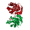
- PDB-9fkc: Crystal structure of human Glucose-6-phosphate isomerase with cit... -
+ Open data

- Basic information
| Entry | Database: PDB / ID: 9fkc | |||||||||
|---|---|---|---|---|---|---|---|---|---|---|
| Title | Crystal structure of human Glucose-6-phosphate isomerase with citraconate ligand | |||||||||
 Components Components | Glucose-6-phosphate isomerase | |||||||||
 Keywords Keywords | ISOMERASE / Metabolic regulation / glycolysis / modular inhibition | |||||||||
| Function / homology |  Function and homology information Function and homology informationIsomerases; Racemases and epimerases; Acting on carbohydrates and derivatives / racemase and epimerase activity, acting on carbohydrates and derivatives / glucose-6-phosphate isomerase / glucose-6-phosphate isomerase activity / hemostasis / glucose 6-phosphate metabolic process / carbohydrate derivative binding / fructose 6-phosphate metabolic process / monosaccharide binding / Gluconeogenesis ...Isomerases; Racemases and epimerases; Acting on carbohydrates and derivatives / racemase and epimerase activity, acting on carbohydrates and derivatives / glucose-6-phosphate isomerase / glucose-6-phosphate isomerase activity / hemostasis / glucose 6-phosphate metabolic process / carbohydrate derivative binding / fructose 6-phosphate metabolic process / monosaccharide binding / Gluconeogenesis / Glycolysis / canonical glycolysis / ciliary membrane / positive regulation of immunoglobulin production / response to testosterone / erythrocyte homeostasis / negative regulation of glycolytic process through fructose-6-phosphate / response to immobilization stress / humoral immune response / mesoderm formation / response to cadmium ion / response to muscle stretch / response to progesterone / positive regulation of endothelial cell migration / cytokine activity / glycolytic process / gluconeogenesis / TP53 Regulates Metabolic Genes / growth factor activity / response to estradiol / glucose homeostasis / secretory granule lumen / carbohydrate metabolic process / in utero embryonic development / ficolin-1-rich granule lumen / learning or memory / Neutrophil degranulation / ubiquitin protein ligase binding / negative regulation of apoptotic process / extracellular exosome / extracellular region / membrane / cytosol Similarity search - Function | |||||||||
| Biological species |  Homo sapiens (human) Homo sapiens (human) | |||||||||
| Method |  X-RAY DIFFRACTION / SYNCHROTRON / MOLECULAR REPLACEMENT / Resolution: 1.6 Å X-RAY DIFFRACTION / SYNCHROTRON / MOLECULAR REPLACEMENT / Resolution: 1.6 Å | |||||||||
 Authors Authors | Jonatansdottir, Y.Y. / Hjorleifsson, G.J. | |||||||||
| Funding support |  Iceland, 2items Iceland, 2items
| |||||||||
 Citation Citation | Journal: Febs J. / Year: 2025 Title: Human glycolysis isomerases are inhibited by weak metabolite modulators. Authors: Jonatansdottir, Y.Y. / Rolfsson, O. / Hjorleifsson, J.G. | |||||||||
| History |
|
- Structure visualization
| Structure viewer | Molecule: MolmilJmol/JSmol |
|---|
- Downloads & links
-Download
| PDBx/mmCIF format | 9fkc.cif.gz | 1 MB | Display | PDBx/mmCIF format |
|---|---|---|---|---|
| PDB format | pdb9fkc.ent.gz | 713.5 KB | Display | PDB format |
| PDBx/mmJSON format | 9fkc.json.gz | Tree view | PDBx/mmJSON format | |
| Others |  Other downloads Other downloads |
-Validation report
| Arichive directory | https://data.pdbj.org/pub/pdb/validation_reports/fk/9fkcftp://data.pdbj.org/pub/pdb/validation_reports/fk/9fkc | HTTPS FTP |
|---|
-Related structure data


-Links
 PDBj
PDBj


- Assembly
| Deposited unit | 
| ||||||||||||
|---|---|---|---|---|---|---|---|---|---|---|---|---|---|
| 1 | 
| ||||||||||||
| 2 | 
| ||||||||||||
| Unit cell |
|
-Components
-Protein , 1 types, 4 molecules ABCD
| #1: Protein | Mass: 63229.949 Da / Num. of mol.: 4 Source method: isolated from a genetically manipulated source Source: (gene. exp.) Homo sapiens (human) / Gene: GPI / Production host:  |
|---|
-Non-polymers , 6 types, 1755 molecules 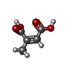


| #2: Chemical | ChemComp-CIZ / (~{  Mass: 130.099 Da / Num. of mol.: 4 / Source method: obtained synthetically / Formula: C5H6O4 / Feature type: SUBJECT OF INVESTIGATION Mass: 130.099 Da / Num. of mol.: 4 / Source method: obtained synthetically / Formula: C5H6O4 / Feature type: SUBJECT OF INVESTIGATION#3: Chemical |  Mass: 238.278 Da / Num. of mol.: 2 / Source method: obtained synthetically / Formula: C10H22O6 / Comment: precipitant*YM Mass: 238.278 Da / Num. of mol.: 2 / Source method: obtained synthetically / Formula: C10H22O6 / Comment: precipitant*YM#4: Chemical | ChemComp-PG4 / |  Mass: 194.226 Da / Num. of mol.: 1 / Source method: isolated from a natural source / Formula: C8H18O5 / Comment: precipitant*YM Mass: 194.226 Da / Num. of mol.: 1 / Source method: isolated from a natural source / Formula: C8H18O5 / Comment: precipitant*YM#5: Chemical |  Mass: 150.173 Da / Num. of mol.: 2 / Source method: obtained synthetically / Formula: C6H14O4 Mass: 150.173 Da / Num. of mol.: 2 / Source method: obtained synthetically / Formula: C6H14O4#6: Chemical | ChemComp-BME / |  Mass: 78.133 Da / Num. of mol.: 1 / Source method: obtained synthetically / Formula: C2H6OS Mass: 78.133 Da / Num. of mol.: 1 / Source method: obtained synthetically / Formula: C2H6OS#7: Water | ChemComp-HOH / | Mass: 18.015 Da / Num. of mol.: 1745 / Source method: isolated from a natural source / Formula: H2O |
|---|
-Details
| Has ligand of interest | Y |
|---|---|
| Has protein modification | N |
-Experimental details
-Experiment
| Experiment | Method: X-RAY DIFFRACTION / Number of used crystals: 1 |
|---|
- Sample preparation
| Crystal | Density Matthews: 2.34 Å3/Da / Density % sol: 47.35 % |
|---|---|
| Crystal grow | Temperature: 295 K / Method: vapor diffusion, hanging drop / pH: 7 Details: Protein buffer: 20 mM Tris pH 7.4, 30 mM NaCl, 20 mM citraconate ligand. Reservoir: 21% w/v PEG3500, 0.16 M CaCl2, and 0.058 M HEPES, pH 7.0 Co-crystallization with ligand: Hanging drop: 1.5: ...Details: Protein buffer: 20 mM Tris pH 7.4, 30 mM NaCl, 20 mM citraconate ligand. Reservoir: 21% w/v PEG3500, 0.16 M CaCl2, and 0.058 M HEPES, pH 7.0 Co-crystallization with ligand: Hanging drop: 1.5:0.5:1.5 ul - Protein (8 mg/ml):Seed stock:Reservoir. Cryoprotectant = a mixture containing the mother liquor, 24% v/v glycerol, and 15-20 mM ligand. |
-Data collection
| Diffraction | Mean temperature: 100 K / Serial crystal experiment: N |
|---|---|
| Diffraction source | Source: SYNCHROTRON / Site: MAX IV  / Beamline: BioMAX / Wavelength: 0.987 Å / Beamline: BioMAX / Wavelength: 0.987 Å |
| Detector | Type: DECTRIS EIGER X 16M / Detector: PIXEL / Date: Dec 12, 2023 |
| Radiation | Protocol: SINGLE WAVELENGTH / Monochromatic (M) / Laue (L): M / Scattering type: x-ray |
| Radiation wavelength | Wavelength: 0.987 Å / Relative weight: 1 |
| Reflection | Resolution: 1.6→46.32 Å / Num. obs: 310523 / % possible obs: 99.67 % / Redundancy: 9.1 % / Biso Wilson estimate: 21.02 Å2 / CC1/2: 0.998 / CC star: 1 / Rmerge(I) obs: 0.1769 / Rpim(I) all: 0.06094 / Rrim(I) all: 0.1873 / Net I/σ(I): 8.66 |
| Reflection shell | Resolution: 1.6→1.657 Å / Redundancy: 9.2 % / Rmerge(I) obs: 3.608 / Mean I/σ(I) obs: 0.93 / Num. unique obs: 30661 / CC1/2: 0.464 / CC star: 0.796 / Rpim(I) all: 1.232 / Rrim(I) all: 3.816 / % possible all: 99.1 |
- Processing
| Software |
| |||||||||||||||||||||||||||||||||||||||||||||||||||||||||||||||||||||||||||||||||||||||||||||||||||||||||||||||||||||||||||||||||||||||||||||||||||||||||||||||||||||||||||||||||||||||||||||||||||||||||||||||||||||||||
|---|---|---|---|---|---|---|---|---|---|---|---|---|---|---|---|---|---|---|---|---|---|---|---|---|---|---|---|---|---|---|---|---|---|---|---|---|---|---|---|---|---|---|---|---|---|---|---|---|---|---|---|---|---|---|---|---|---|---|---|---|---|---|---|---|---|---|---|---|---|---|---|---|---|---|---|---|---|---|---|---|---|---|---|---|---|---|---|---|---|---|---|---|---|---|---|---|---|---|---|---|---|---|---|---|---|---|---|---|---|---|---|---|---|---|---|---|---|---|---|---|---|---|---|---|---|---|---|---|---|---|---|---|---|---|---|---|---|---|---|---|---|---|---|---|---|---|---|---|---|---|---|---|---|---|---|---|---|---|---|---|---|---|---|---|---|---|---|---|---|---|---|---|---|---|---|---|---|---|---|---|---|---|---|---|---|---|---|---|---|---|---|---|---|---|---|---|---|---|---|---|---|---|---|---|---|---|---|---|---|---|---|---|---|---|---|---|---|---|
| Refinement | Method to determine structure: MOLECULAR REPLACEMENT / Resolution: 1.6→46.32 Å / SU ML: 0.2238 / Cross valid method: FREE R-VALUE / σ(F): 1.33 / Phase error: 28.2535 Stereochemistry target values: GeoStd + Monomer Library + CDL v1.2
| |||||||||||||||||||||||||||||||||||||||||||||||||||||||||||||||||||||||||||||||||||||||||||||||||||||||||||||||||||||||||||||||||||||||||||||||||||||||||||||||||||||||||||||||||||||||||||||||||||||||||||||||||||||||||
| Solvent computation | Shrinkage radii: 0.9 Å / VDW probe radii: 1.1 Å / Solvent model: FLAT BULK SOLVENT MODEL | |||||||||||||||||||||||||||||||||||||||||||||||||||||||||||||||||||||||||||||||||||||||||||||||||||||||||||||||||||||||||||||||||||||||||||||||||||||||||||||||||||||||||||||||||||||||||||||||||||||||||||||||||||||||||
| Displacement parameters | Biso mean: 29.96 Å2 | |||||||||||||||||||||||||||||||||||||||||||||||||||||||||||||||||||||||||||||||||||||||||||||||||||||||||||||||||||||||||||||||||||||||||||||||||||||||||||||||||||||||||||||||||||||||||||||||||||||||||||||||||||||||||
| Refinement step | Cycle: LAST / Resolution: 1.6→46.32 Å
| |||||||||||||||||||||||||||||||||||||||||||||||||||||||||||||||||||||||||||||||||||||||||||||||||||||||||||||||||||||||||||||||||||||||||||||||||||||||||||||||||||||||||||||||||||||||||||||||||||||||||||||||||||||||||
| Refine LS restraints |
| |||||||||||||||||||||||||||||||||||||||||||||||||||||||||||||||||||||||||||||||||||||||||||||||||||||||||||||||||||||||||||||||||||||||||||||||||||||||||||||||||||||||||||||||||||||||||||||||||||||||||||||||||||||||||
| LS refinement shell |
|
