 Movie
Movie Controller


+ Open data

- Basic information
| Entry | Database: PDB / ID: 9fh0 | ||||||||||||||||||||||||||||||
|---|---|---|---|---|---|---|---|---|---|---|---|---|---|---|---|---|---|---|---|---|---|---|---|---|---|---|---|---|---|---|---|
| Title | Pex5-Eci1 complex - Pex5 local refinement | ||||||||||||||||||||||||||||||
 Components Components |
| ||||||||||||||||||||||||||||||
 Keywords Keywords | PROTEIN TRANSPORT / Peroxisome / protein targeting / PTS1 | ||||||||||||||||||||||||||||||
| Function / homology |  Function and homology information Function and homology informationBeta-oxidation of very long chain fatty acids / Pexophagy / peroxisome matrix targeting signal-1 binding / peroxisomal importomer complex / protein import into peroxisome matrix / Peroxisomal protein import / protein import into peroxisome matrix, docking / Delta3-Delta2-enoyl-CoA isomerase / delta(3)-delta(2)-enoyl-CoA isomerase activity / protein carrier activity ...Beta-oxidation of very long chain fatty acids / Pexophagy / peroxisome matrix targeting signal-1 binding / peroxisomal importomer complex / protein import into peroxisome matrix / Peroxisomal protein import / protein import into peroxisome matrix, docking / Delta3-Delta2-enoyl-CoA isomerase / delta(3)-delta(2)-enoyl-CoA isomerase activity / protein carrier activity / peroxisomal membrane / fatty acid beta-oxidation / peroxisomal matrix / peroxisome / protein-macromolecule adaptor activity / cytosol Similarity search - Function | ||||||||||||||||||||||||||||||
| Biological species |  | ||||||||||||||||||||||||||||||
| Method | ELECTRON MICROSCOPY / single particle reconstruction / cryo EM / Resolution: 2.9 Å | ||||||||||||||||||||||||||||||
 Authors Authors | Elad, N. / Dym, O. | ||||||||||||||||||||||||||||||
| Funding support | European Union,  Israel, 2items Israel, 2items
| ||||||||||||||||||||||||||||||
 Citation Citation | Journal: J Cell Sci / Year: 2025 Title: A cryo-electron microscopy structure of yeast Pex5 in complex with a cargo uncovers a novel binding interface. Authors: Lior Peer / Orly Dym / Nadav Elad / Asa Tirosh / Jossef Jacobovitch / Ehud Sivan / Mor Angel / Shira Albeck / Maya Schuldiner / Yoav Peleg / Einat Zalckvar / Abstract: Proper protein targeting to organelles is crucial for maintaining eukaryotic cellular function and homeostasis. This necessity has driven the evolution of specific targeting signals on proteins and ...Proper protein targeting to organelles is crucial for maintaining eukaryotic cellular function and homeostasis. This necessity has driven the evolution of specific targeting signals on proteins and the targeting factors that recognize them. A prominent example is peroxisomal matrix proteins, most of which depend on the targeting factor Pex5 to localize and function correctly. Although most Pex5 cargoes contain a peroxisomal targeting signal type 1 (PTS1), they are not all targeted similarly. Some undergo priority targeting, facilitated either by stronger binding to specific subsets of PTS1 signals or by additional interaction interfaces. These observations highlight the extensive complexity of Pex5-mediated targeting. In this study, we reveal that the Saccharomyces cerevisiae (yeast) matrix protein Eci1 can reach peroxisomes and bind Pex5 in the absence of PTS1. By solving the structure of the yeast Pex5-Eci1 complex using cryo-electron microscopy, we identified additional binding interfaces. Our findings provide new insights into the versatile interactions between Pex5 and its cargo, Eci1. More broadly, this work highlights the intricate, dynamic nature of the interactions between cargo factors and their cargoes to meet the complex environment within eukaryotic cells. | ||||||||||||||||||||||||||||||
| History |
|
- Structure visualization
| Structure viewer | Molecule: MolmilJmol/JSmol |
|---|
- Downloads & links
-Download
| PDBx/mmCIF format | 9fh0.cif.gz | 118.5 KB | Display | PDBx/mmCIF format |
|---|---|---|---|---|
| PDB format | pdb9fh0.ent.gz | 85.9 KB | Display | PDB format |
| PDBx/mmJSON format | 9fh0.json.gz | Tree view | PDBx/mmJSON format | |
| Others |  Other downloads Other downloads |
-Validation report
| Arichive directory | https://data.pdbj.org/pub/pdb/validation_reports/fh/9fh0ftp://data.pdbj.org/pub/pdb/validation_reports/fh/9fh0 | HTTPS FTP |
|---|
-Related structure data
| Related structure data | 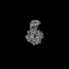 50435MC  9fgzC M: map data used to model this data C: citing same article ( |
|---|---|
| Similar structure data |

-Links
 PDBj
PDBj


- Assembly
| Deposited unit | 
|
|---|---|
| 1 |
|
-Components
| #1: Protein | Mass: 31736.408 Da / Num. of mol.: 1 Source method: isolated from a genetically manipulated source Source: (gene. exp.) Gene: ECI1 / Production host:  |
|---|---|
| #2: Protein | Mass: 69335.766 Da / Num. of mol.: 1 Source method: isolated from a genetically manipulated source Source: (gene. exp.) Gene: PEX5, PAS10, YDR244W, YD8419.11 / Production host:  Escherichia phage EcSzw-2 (virus) / References: UniProt: P35056 Escherichia phage EcSzw-2 (virus) / References: UniProt: P35056 |
| Has protein modification | N |
-Experimental details
-Experiment
| Experiment | Method: ELECTRON MICROSCOPY |
|---|---|
| EM experiment | Aggregation state: PARTICLE / 3D reconstruction method: single particle reconstruction |
- Sample preparation
| Component | Name: Pex5-Eci1 complex / Type: COMPLEX / Entity ID: all / Source: RECOMBINANT |
|---|---|
| Source (natural) | Organism: |
| Source (recombinant) | Organism: |
| Buffer solution | pH: 7.5 |
| Specimen | Embedding applied: NO / Shadowing applied: NO / Staining applied: NO / Vitrification applied: YES |
| Specimen support | Grid material: GOLD / Grid mesh size: 300 divisions/in. / Grid type: C-flat-1.2/1.3 |
| Vitrification | Instrument: FEI VITROBOT MARK IV / Cryogen name: ETHANE / Humidity: 100 % / Chamber temperature: 277 K |
- Electron microscopy imaging
| Experimental equipment |  Model: Titan Krios / Image courtesy: FEI Company |
|---|---|
| Microscopy | Model: FEI TITAN KRIOS |
| Electron gun | Electron source:  FIELD EMISSION GUN / Accelerating voltage: 300 kV / Illumination mode: FLOOD BEAM FIELD EMISSION GUN / Accelerating voltage: 300 kV / Illumination mode: FLOOD BEAM |
| Electron lens | Mode: BRIGHT FIELD / Nominal magnification: 105000 X / Calibrated magnification: 59382 X / Nominal defocus max: 15000 nm / Nominal defocus min: 2700 nm / Cs: 2.7 mm / C2 aperture diameter: 70 µm / Alignment procedure: COMA FREE |
| Specimen holder | Cryogen: NITROGEN / Specimen holder model: FEI TITAN KRIOS AUTOGRID HOLDER |
| Image recording | Average exposure time: 1.6 sec. / Electron dose: 45.5 e/Å2 / Film or detector model: GATAN K3 BIOQUANTUM (6k x 4k) / Num. of grids imaged: 1 / Num. of real images: 5577 |
| EM imaging optics | Energyfilter name: GIF Bioquantum / Energyfilter slit width: 15 eV |
- Processing
| EM software |
| |||||||||||||||||||||||||||||||||||||||||||||
|---|---|---|---|---|---|---|---|---|---|---|---|---|---|---|---|---|---|---|---|---|---|---|---|---|---|---|---|---|---|---|---|---|---|---|---|---|---|---|---|---|---|---|---|---|---|---|
| CTF correction | Type: PHASE FLIPPING AND AMPLITUDE CORRECTION | |||||||||||||||||||||||||||||||||||||||||||||
| Particle selection | Num. of particles selected: 2170575 | |||||||||||||||||||||||||||||||||||||||||||||
| Symmetry | Point symmetry: C1 (asymmetric) | |||||||||||||||||||||||||||||||||||||||||||||
| 3D reconstruction | Resolution: 2.9 Å / Resolution method: FSC 0.143 CUT-OFF / Num. of particles: 117945 / Symmetry type: POINT | |||||||||||||||||||||||||||||||||||||||||||||
| Refine LS restraints |
|
