+ Open data
- Basic information
| Entry | Database: PDB / ID: 9fgn | ||||||||||||
|---|---|---|---|---|---|---|---|---|---|---|---|---|---|
| Title | Coxsackievirus A9 bound with compound 18 (CL304) | ||||||||||||
 Components Components |
| ||||||||||||
 Keywords Keywords | VIRUS / Antiviral / capsid stabilizer / hydrophobic pocket / cryoEM | ||||||||||||
| Function / homology |  Function and homology information Function and homology informationsymbiont-mediated suppression of host cytoplasmic pattern recognition receptor signaling pathway via inhibition of RIG-I activity / picornain 2A / symbiont-mediated suppression of host mRNA export from nucleus / symbiont genome entry into host cell via pore formation in plasma membrane / picornain 3C / T=pseudo3 icosahedral viral capsid / host cell cytoplasmic vesicle membrane / nucleoside-triphosphate phosphatase / channel activity / monoatomic ion transmembrane transport ...symbiont-mediated suppression of host cytoplasmic pattern recognition receptor signaling pathway via inhibition of RIG-I activity / picornain 2A / symbiont-mediated suppression of host mRNA export from nucleus / symbiont genome entry into host cell via pore formation in plasma membrane / picornain 3C / T=pseudo3 icosahedral viral capsid / host cell cytoplasmic vesicle membrane / nucleoside-triphosphate phosphatase / channel activity / monoatomic ion transmembrane transport / DNA replication / RNA helicase activity / endocytosis involved in viral entry into host cell / symbiont-mediated activation of host autophagy / RNA-directed RNA polymerase / cysteine-type endopeptidase activity / viral RNA genome replication / RNA-directed RNA polymerase activity / DNA-templated transcription / virion attachment to host cell / host cell nucleus / structural molecule activity / ATP hydrolysis activity / proteolysis / RNA binding / zinc ion binding / ATP binding / membrane Similarity search - Function | ||||||||||||
| Biological species |  Coxsackievirus A9 Coxsackievirus A9 | ||||||||||||
| Method | ELECTRON MICROSCOPY / single particle reconstruction / cryo EM / Resolution: 2.64 Å | ||||||||||||
 Authors Authors | Plavec, Z. / Butcher, S.J. / Mitchell, C. / Buckner, C. | ||||||||||||
| Funding support |  Finland, 3items Finland, 3items
| ||||||||||||
 Citation Citation |  Journal: J Med Chem / Year: 2024 Journal: J Med Chem / Year: 2024Title: SAR Analysis of Novel Coxsackie virus A9 Capsid Binders. Authors: Chiara Tammaro / Zlatka Plavec / Laura Myllymäki / Cristopher Mitchell / Sara Consalvi / Mariangela Biava / Alessia Ciogli / Aušra Domanska / Valtteri Leppilampi / Cienna Buckner / Simone ...Authors: Chiara Tammaro / Zlatka Plavec / Laura Myllymäki / Cristopher Mitchell / Sara Consalvi / Mariangela Biava / Alessia Ciogli / Aušra Domanska / Valtteri Leppilampi / Cienna Buckner / Simone Manetto / Pietro Sciò / Antonio Coluccia / Mira Laajala / Giulio M Dondio / Chiara Bigogno / Varpu Marjomäki / Sarah J Butcher / Giovanna Poce /   Abstract: Enterovirus infections are common in humans, yet there are no approved antiviral treatments. In this study we concentrated on inhibition of one of the (EV-B), namely Coxsackievirus A9 (CVA9), using ...Enterovirus infections are common in humans, yet there are no approved antiviral treatments. In this study we concentrated on inhibition of one of the (EV-B), namely Coxsackievirus A9 (CVA9), using a combination of medicinal chemistry, virus inhibition assays, structure determination from cryogenic electron microscopy and molecular modeling, to determine the structure activity relationships for a promising class of novel -phenylbenzylamines. Of the new 29 compounds synthesized, 10 had half maximal effective concentration (EC) values between 0.64-10.46 μM, and of these, 7 had 50% cytotoxicity concentration (CC) values higher than 200 μM. In addition, this new series of compounds showed promising physicochemical properties and act through capsid stabilization, preventing capsid expansion and subsequent release of the genome. #1: Journal: Protein Sci / Year: 2021 Title: UCSF ChimeraX: Structure visualization for researchers, educators, and developers. Authors: Eric F Pettersen / Thomas D Goddard / Conrad C Huang / Elaine C Meng / Gregory S Couch / Tristan I Croll / John H Morris / Thomas E Ferrin /   Abstract: UCSF ChimeraX is the next-generation interactive visualization program from the Resource for Biocomputing, Visualization, and Informatics (RBVI), following UCSF Chimera. ChimeraX brings (a) ...UCSF ChimeraX is the next-generation interactive visualization program from the Resource for Biocomputing, Visualization, and Informatics (RBVI), following UCSF Chimera. ChimeraX brings (a) significant performance and graphics enhancements; (b) new implementations of Chimera's most highly used tools, many with further improvements; (c) several entirely new analysis features; (d) support for new areas such as virtual reality, light-sheet microscopy, and medical imaging data; (e) major ease-of-use advances, including toolbars with icons to perform actions with a single click, basic "undo" capabilities, and more logical and consistent commands; and (f) an app store for researchers to contribute new tools. ChimeraX includes full user documentation and is free for noncommercial use, with downloads available for Windows, Linux, and macOS from https://www.rbvi.ucsf.edu/chimerax. | ||||||||||||
| History |
|
- Structure visualization
| Structure viewer | Molecule:  Molmil Molmil Jmol/JSmol Jmol/JSmol |
|---|
- Downloads & links
- Download
| PDBx/mmCIF format |  9fgn.cif.gz 9fgn.cif.gz | 278.4 KB | Display |  PDBx/mmCIF format PDBx/mmCIF format |
|---|---|---|---|---|
| PDB format |  pdb9fgn.ent.gz pdb9fgn.ent.gz | Display |  PDB format PDB format | |
| PDBx/mmJSON format |  9fgn.json.gz 9fgn.json.gz | Tree view |  PDBx/mmJSON format PDBx/mmJSON format | |
| Others |  Other downloads Other downloads |
-Validation report
| Arichive directory |  https://data.pdbj.org/pub/pdb/validation_reports/fg/9fgn https://data.pdbj.org/pub/pdb/validation_reports/fg/9fgn ftp://data.pdbj.org/pub/pdb/validation_reports/fg/9fgn ftp://data.pdbj.org/pub/pdb/validation_reports/fg/9fgn | HTTPS FTP |
|---|
-Related structure data
| Related structure data |  50414MC 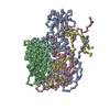 8s7jC  9exiC  9fa9C  9fczC  9fo2C  9fo5C  9fp5C C: citing same article ( M: map data used to model this data |
|---|---|
| Similar structure data | Similarity search - Function & homology  F&H Search F&H Search |
- Links
Links
- Assembly
| Deposited unit | 
|
|---|---|
| 1 | x 60
|
- Components
| #1: Protein | Mass: 31952.896 Da / Num. of mol.: 1 / Source method: isolated from a natural source / Source: (natural)  Coxsackievirus A9 / Cell line: GMK / Strain: Griggs / Tissue: kidney / References: UniProt: P21404 Coxsackievirus A9 / Cell line: GMK / Strain: Griggs / Tissue: kidney / References: UniProt: P21404 |
|---|---|
| #2: Protein | Mass: 27720.285 Da / Num. of mol.: 1 / Source method: isolated from a natural source / Source: (natural)  Coxsackievirus A9 / Cell line: GMK / Strain: Griggs / Tissue: kidney / References: UniProt: P21404 Coxsackievirus A9 / Cell line: GMK / Strain: Griggs / Tissue: kidney / References: UniProt: P21404 |
| #3: Protein | Mass: 26149.889 Da / Num. of mol.: 1 / Source method: isolated from a natural source / Source: (natural)  Coxsackievirus A9 / Cell line: GMK / Strain: Griggs / Tissue: kidney / References: UniProt: P21404 Coxsackievirus A9 / Cell line: GMK / Strain: Griggs / Tissue: kidney / References: UniProt: P21404 |
| #4: Protein | Mass: 7237.936 Da / Num. of mol.: 1 / Source method: isolated from a natural source / Source: (natural)  Coxsackievirus A9 / Cell line: GMK / Strain: Griggs / Tissue: kidney / References: UniProt: P21404 Coxsackievirus A9 / Cell line: GMK / Strain: Griggs / Tissue: kidney / References: UniProt: P21404 |
| #5: Chemical | ChemComp-A1ICH / ~{ Mass: 331.403 Da / Num. of mol.: 1 / Source method: obtained synthetically / Formula: C19H23F2N3 |
| Has protein modification | N |
-Experimental details
-Experiment
| Experiment | Method: ELECTRON MICROSCOPY |
|---|---|
| EM experiment | Aggregation state: PARTICLE / 3D reconstruction method: single particle reconstruction |
- Sample preparation
| Component | Name: Human coxsackievirus A9 (strain Griggs) / Type: VIRUS Details: Coxsackievirus A9 was propagated on green monkey kidney cells and purified on a sucrose gradient. Entity ID: #1-#4 / Source: NATURAL |
|---|---|
| Molecular weight | Value: 8 MDa / Experimental value: NO |
| Source (natural) | Organism:  Human coxsackievirus A9 (strain Griggs) / Strain: Griggs Human coxsackievirus A9 (strain Griggs) / Strain: Griggs |
| Details of virus | Empty: NO / Enveloped: NO / Isolate: STRAIN / Type: VIRION |
| Natural host | Organism: Homo sapiens |
| Virus shell | Name: icosahedral capsid / Diameter: 300 nm / Triangulation number (T number): 1 |
| Buffer solution | pH: 7.2 / Details: PBS containing 2 mM MgCl2 and 5% DMSO. |
| Specimen | Conc.: 0.4 mg/ml / Embedding applied: NO / Shadowing applied: NO / Staining applied: NO / Vitrification applied: YES Details: Compound 18 (CL304) was solubilized in DMSO and diluted to the final concentration in PBS + 2 mM MgCl2. Purified Coxsackievirus A9 was incubated with compound 18 (CL304) for 1h at 37C after ...Details: Compound 18 (CL304) was solubilized in DMSO and diluted to the final concentration in PBS + 2 mM MgCl2. Purified Coxsackievirus A9 was incubated with compound 18 (CL304) for 1h at 37C after which the sample was vitrified on a semi-automatic plunger Leica EM GP on copper Quantifoil R1.2/1.3 grids with 2 nm thin carbon film and 300 mesh. This sample was monodisperse. |
| Specimen support | Grid material: COPPER / Grid mesh size: 300 divisions/in. / Grid type: Quantifoil R1.2/1.3 |
| Vitrification | Instrument: LEICA EM GP / Cryogen name: ETHANE / Humidity: 80 % / Chamber temperature: 295 K Details: Sample was incubated for 15 s on the grid before blotted from the front for 1.5 s. |
- Electron microscopy imaging
| Experimental equipment |  Model: Talos Arctica / Image courtesy: FEI Company |
|---|---|
| Microscopy | Model: FEI TALOS ARCTICA |
| Electron gun | Electron source:  FIELD EMISSION GUN / Accelerating voltage: 200 kV / Illumination mode: FLOOD BEAM FIELD EMISSION GUN / Accelerating voltage: 200 kV / Illumination mode: FLOOD BEAM |
| Electron lens | Mode: BRIGHT FIELD / Nominal magnification: 150000 X / Nominal defocus max: 2300 nm / Nominal defocus min: 600 nm |
| Specimen holder | Cryogen: NITROGEN / Temperature (max): 103.15 K / Temperature (min): 93.15 K |
| Image recording | Average exposure time: 1 sec. / Electron dose: 40 e/Å2 / Detector mode: COUNTING / Film or detector model: FEI FALCON III (4k x 4k) / Num. of real images: 625 |
| Image scans | Width: 1496 / Height: 1496 |
- Processing
| EM software |
|
|---|
 Movie
Movie Controller


 PDBj
PDBj
