 Movie
Movie Controller

[English] 日本語
 Yorodumi
- PDB-9fdf: Human phosphoglycerate kinase in with mixture of products and sub... -
+ Open data

- Basic information
| Entry | Database: PDB / ID: 9fdf | ||||||
|---|---|---|---|---|---|---|---|
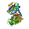
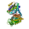
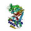
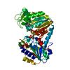
| Title | Human phosphoglycerate kinase in with mixture of products and substrates produced by cross-soaking a TSA crystal | ||||||
 Components Components | Phosphoglycerate kinase 1 | ||||||
 Keywords Keywords | TRANSFERASE / Phosphoryl transfer Glycolysis ATP binding Rossmann fold | ||||||
| Function / homology |  Function and homology information Function and homology informationnegative regulation of pyruvate decarboxylation to acetyl-CoA / Manipulation of host energy metabolism / phosphoglycerate kinase / phosphoglycerate kinase activity / protein-disulfide reductase [NAD(P)H] activity / Gluconeogenesis / Glycolysis / plasminogen activation / canonical glycolysis / epithelial cell differentiation ...negative regulation of pyruvate decarboxylation to acetyl-CoA / Manipulation of host energy metabolism / phosphoglycerate kinase / phosphoglycerate kinase activity / protein-disulfide reductase [NAD(P)H] activity / Gluconeogenesis / Glycolysis / plasminogen activation / canonical glycolysis / epithelial cell differentiation / negative regulation of angiogenesis / glycolytic process / gluconeogenesis / ADP binding / cellular response to hypoxia / transmembrane transporter binding / non-specific serine/threonine protein kinase / membrane raft / mitochondrial matrix / protein serine kinase activity / protein serine/threonine kinase activity / : / extracellular exosome / ATP binding / membrane / metal ion binding / cytosol Similarity search - Function | ||||||
| Biological species |  Homo sapiens (human) Homo sapiens (human) | ||||||
| Method |  X-RAY DIFFRACTION / SYNCHROTRON / MOLECULAR REPLACEMENT / Resolution: 1.44 Å X-RAY DIFFRACTION / SYNCHROTRON / MOLECULAR REPLACEMENT / Resolution: 1.44 Å | ||||||
 Authors Authors | Cliff, M.J. / Waltho, J.P. / Bowler, M.W. / Baxter, N.J. / Bisson, C. / Blackburn, G.M. | ||||||
| Funding support |  United Kingdom, 1items United Kingdom, 1items
| ||||||
 Citation Citation | Journal: To be published Title: The role of magnesium in catalysis by phosphoglycerate kinase Authors: Cliff, M.J. / Serimbetov, Z. / Bisson, C. / Baxter, N.J. / Blackburn, G.M. / Hay, S. / Bowler, M.W. / Waltho, J.P. | ||||||
| History |
|
- Structure visualization
| Structure viewer | Molecule: MolmilJmol/JSmol |
|---|
- Downloads & links
-Download
| PDBx/mmCIF format | 9fdf.cif.gz | 310 KB | Display | PDBx/mmCIF format |
|---|---|---|---|---|
| PDB format | pdb9fdf.ent.gz | 206.4 KB | Display | PDB format |
| PDBx/mmJSON format | 9fdf.json.gz | Tree view | PDBx/mmJSON format | |
| Others |  Other downloads Other downloads |
-Validation report
| Arichive directory | https://data.pdbj.org/pub/pdb/validation_reports/fd/9fdfftp://data.pdbj.org/pub/pdb/validation_reports/fd/9fdf | HTTPS FTP |
|---|
-Related structure data


-Links
 PDBj
PDBj

- Assembly
| Deposited unit | 
| ||||||||||||
|---|---|---|---|---|---|---|---|---|---|---|---|---|---|
| 1 |
| ||||||||||||
| Unit cell |
|
-Components
-Protein , 1 types, 1 molecules A
| #1: Protein | Mass: 44672.621 Da / Num. of mol.: 1 Source method: isolated from a genetically manipulated source Source: (gene. exp.) Homo sapiens (human) / Gene: PGK1, PGKA, MIG10, OK/SW-cl.110 / Production host:  |
|---|
-Non-polymers , 5 types, 523 molecules 


| #2: Chemical | ChemComp-ATP /  Mass: 507.181 Da / Num. of mol.: 1 / Source method: obtained synthetically / Formula: C10H16N5O13P3 / Feature type: SUBJECT OF INVESTIGATION / Comment: ATP, energy-carrying molecule*YM Mass: 507.181 Da / Num. of mol.: 1 / Source method: obtained synthetically / Formula: C10H16N5O13P3 / Feature type: SUBJECT OF INVESTIGATION / Comment: ATP, energy-carrying molecule*YM |
|---|---|
| #3: Chemical | ChemComp-X15 /  Mass: 266.037 Da / Num. of mol.: 1 / Source method: obtained synthetically / Formula: C3H8O10P2 / Feature type: SUBJECT OF INVESTIGATION Mass: 266.037 Da / Num. of mol.: 1 / Source method: obtained synthetically / Formula: C3H8O10P2 / Feature type: SUBJECT OF INVESTIGATION |
| #4: Chemical | ChemComp-NA /  Mass: 22.990 Da / Num. of mol.: 1 / Source method: obtained synthetically / Formula: Na / Feature type: SUBJECT OF INVESTIGATION Mass: 22.990 Da / Num. of mol.: 1 / Source method: obtained synthetically / Formula: Na / Feature type: SUBJECT OF INVESTIGATION |
| #5: Chemical | ChemComp-CL /  Mass: 35.453 Da / Num. of mol.: 1 / Source method: obtained synthetically / Formula: Cl Mass: 35.453 Da / Num. of mol.: 1 / Source method: obtained synthetically / Formula: Cl |
| #6: Water | ChemComp-HOH / Mass: 18.015 Da / Num. of mol.: 519 / Source method: isolated from a natural source / Formula: H2O |
-Details
| Has ligand of interest | Y |
|---|---|
| Has protein modification | N |
-Experimental details
-Experiment
| Experiment | Method: X-RAY DIFFRACTION / Number of used crystals: 1 |
|---|
- Sample preparation
| Crystal | Density Matthews: 2.21 Å3/Da / Density % sol: 44.22 % / Description: plates |
|---|---|
| Crystal grow | Temperature: 298 K / Method: vapor diffusion, sitting drop / pH: 6.5 Details: crystals were initially produced from 50 mM Tris (pH 7.5), 20 mM DTT, 25 mM MgCl2, 50 mM 3PG, and 10 mM ADP, supplemented with 20 mM NH4F and 1 mM deferoxamine; in 28-33% PEG 2000 MME and 0. ...Details: crystals were initially produced from 50 mM Tris (pH 7.5), 20 mM DTT, 25 mM MgCl2, 50 mM 3PG, and 10 mM ADP, supplemented with 20 mM NH4F and 1 mM deferoxamine; in 28-33% PEG 2000 MME and 0.1 M Bis/Tris pH 6.5 then cross-soaked with 35% PEG 2000 MME; 0.1 M Bis/Tris pH 6.5, 20 mM DTT, and 50 mM 3PG and 0.1mM deferoxamine Temp details: "room temperature" |
-Data collection
| Diffraction | Mean temperature: 100 K / Serial crystal experiment: N |
|---|---|
| Diffraction source | Source: SYNCHROTRON / Site: Diamond / Beamline: I04-1 / Wavelength: 0.933 Å |
| Detector | Type: DECTRIS EIGER2 XE 9M / Detector: PIXEL / Date: Jul 23, 2012 |
| Radiation | Protocol: SINGLE WAVELENGTH / Monochromatic (M) / Laue (L): M / Scattering type: x-ray |
| Radiation wavelength | Wavelength: 0.933 Å / Relative weight: 1 |
| Reflection | Resolution: 1.44→37.06 Å / Num. obs: 71262 / % possible obs: 98.48 % / Redundancy: 1.9 % / Biso Wilson estimate: 15.43 Å2 / CC1/2: 0.999 / Rmerge(I) obs: 0.03 / Rpim(I) all: 0.03 / Rrim(I) all: 0.042 / Net I/σ(I): 14.2 |
| Reflection shell | Resolution: 1.44→1.49 Å / Redundancy: 1.9 % / Rmerge(I) obs: 0.353 / Mean I/σ(I) obs: 2.4 / Num. unique obs: 6991 / CC1/2: 0.737 / Rpim(I) all: 0.353 / Rrim(I) all: 0.499 / % possible all: 97.8 |
- Processing
| Software |
| |||||||||||||||||||||||||||||||||||||||||||||||||||||||||||||||||||||||||||||||||||||||||||||||||||||||||||||||||||||||||||||||||||||||||||||||||||||||||||||||||||||||||||||||||||||||||||||
|---|---|---|---|---|---|---|---|---|---|---|---|---|---|---|---|---|---|---|---|---|---|---|---|---|---|---|---|---|---|---|---|---|---|---|---|---|---|---|---|---|---|---|---|---|---|---|---|---|---|---|---|---|---|---|---|---|---|---|---|---|---|---|---|---|---|---|---|---|---|---|---|---|---|---|---|---|---|---|---|---|---|---|---|---|---|---|---|---|---|---|---|---|---|---|---|---|---|---|---|---|---|---|---|---|---|---|---|---|---|---|---|---|---|---|---|---|---|---|---|---|---|---|---|---|---|---|---|---|---|---|---|---|---|---|---|---|---|---|---|---|---|---|---|---|---|---|---|---|---|---|---|---|---|---|---|---|---|---|---|---|---|---|---|---|---|---|---|---|---|---|---|---|---|---|---|---|---|---|---|---|---|---|---|---|---|---|---|---|---|---|
| Refinement | Method to determine structure: MOLECULAR REPLACEMENT / Resolution: 1.44→37.06 Å / SU ML: 0.1353 / Cross valid method: FREE R-VALUE / σ(F): 1.34 / Phase error: 15.6538 Stereochemistry target values: GeoStd + Monomer Library + CDL v1.2
| |||||||||||||||||||||||||||||||||||||||||||||||||||||||||||||||||||||||||||||||||||||||||||||||||||||||||||||||||||||||||||||||||||||||||||||||||||||||||||||||||||||||||||||||||||||||||||||
| Solvent computation | Shrinkage radii: 0.9 Å / VDW probe radii: 1.11 Å / Solvent model: FLAT BULK SOLVENT MODEL | |||||||||||||||||||||||||||||||||||||||||||||||||||||||||||||||||||||||||||||||||||||||||||||||||||||||||||||||||||||||||||||||||||||||||||||||||||||||||||||||||||||||||||||||||||||||||||||
| Displacement parameters | Biso mean: 21.82 Å2 | |||||||||||||||||||||||||||||||||||||||||||||||||||||||||||||||||||||||||||||||||||||||||||||||||||||||||||||||||||||||||||||||||||||||||||||||||||||||||||||||||||||||||||||||||||||||||||||
| Refinement step | Cycle: LAST / Resolution: 1.44→37.06 Å
| |||||||||||||||||||||||||||||||||||||||||||||||||||||||||||||||||||||||||||||||||||||||||||||||||||||||||||||||||||||||||||||||||||||||||||||||||||||||||||||||||||||||||||||||||||||||||||||
| Refine LS restraints |
| |||||||||||||||||||||||||||||||||||||||||||||||||||||||||||||||||||||||||||||||||||||||||||||||||||||||||||||||||||||||||||||||||||||||||||||||||||||||||||||||||||||||||||||||||||||||||||||
| LS refinement shell |
|
