[English] 日本語
Yorodumi
- PDB-9f7w: Humman PPARgamma ligand binding domain in complex with co-activat... -

+
Open data


ID or keywords:

Loading...

-
Basic information

Entry
Database: PDB / ID: 9f7w
TitleHumman PPARgamma ligand binding domain in complex with co-activator 1alpha peptide and bisphenol A (BPA)
Components
  • Peroxisome proliferator-activated receptor gamma
  • Peroxisome proliferator-activated receptor gamma coactivator 1-alpha
KeywordsTRANSCRIPTION / peroxisome proliferator-activated receptor gamma / bisphenol A / NUCLEAR PROTEIN
Function / homology
Function and homology information


: / positive regulation of fatty acid oxidation / prostaglandin receptor activity / regulation of cholesterol transporter activity / negative regulation of connective tissue replacement involved in inflammatory response wound healing / negative regulation of receptor signaling pathway via STAT / MECP2 regulates transcription factors / lncRNA binding / response to muscle activity / negative regulation of extracellular matrix assembly ...: / positive regulation of fatty acid oxidation / prostaglandin receptor activity / regulation of cholesterol transporter activity / negative regulation of connective tissue replacement involved in inflammatory response wound healing / negative regulation of receptor signaling pathway via STAT / MECP2 regulates transcription factors / lncRNA binding / response to muscle activity / negative regulation of extracellular matrix assembly / Activation of PPARGC1A (PGC-1alpha) by phosphorylation / cellular respiration / negative regulation of vascular endothelial cell proliferation / negative regulation of cellular response to transforming growth factor beta stimulus / negative regulation of cardiac muscle hypertrophy in response to stress / arachidonic acid binding / positive regulation of low-density lipoprotein receptor activity / positive regulation of adiponectin secretion / lipoprotein transport / negative regulation of sequestering of triglyceride / DNA binding domain binding / macrophage derived foam cell differentiation / temperature homeostasis / STAT family protein binding / positive regulation of vascular associated smooth muscle cell apoptotic process / positive regulation of fatty acid metabolic process / response to lipid / negative regulation of SMAD protein signal transduction / LBD domain binding / negative regulation of type II interferon-mediated signaling pathway / negative regulation of cholesterol storage / E-box binding / intracellular glucose homeostasis / response to starvation / alpha-actinin binding / fatty acid oxidation / lipid homeostasis / negative regulation of vascular associated smooth muscle cell proliferation / R-SMAD binding / monocyte differentiation / response to dietary excess / negative regulation of macrophage derived foam cell differentiation / negative regulation of blood vessel endothelial cell migration / negative regulation of lipid storage / cellular response to low-density lipoprotein particle stimulus / white fat cell differentiation / negative regulation of BMP signaling pathway / Transcriptional regulation of brown and beige adipocyte differentiation by EBF2 / negative regulation of mitochondrial fission / positive regulation of cholesterol efflux / positive regulation of fat cell differentiation / negative regulation of osteoblast differentiation / retinoic acid receptor signaling pathway / cell fate commitment / adipose tissue development / BMP signaling pathway / FOXO-mediated transcription of oxidative stress, metabolic and neuronal genes / long-chain fatty acid transport / brown fat cell differentiation / energy homeostasis / nuclear retinoid X receptor binding / cell maturation / positive regulation of gluconeogenesis / negative regulation of signaling receptor activity / digestion / epithelial cell differentiation / positive regulation of adipose tissue development / peroxisome proliferator activated receptor signaling pathway / regulation of cellular response to insulin stimulus / hormone-mediated signaling pathway / respiratory electron transport chain / negative regulation of angiogenesis / RNA splicing / response to nutrient / SUMOylation of transcription cofactors / negative regulation of miRNA transcription / mitochondrion organization / nuclear receptor coactivator activity / negative regulation of MAP kinase activity / fatty acid metabolic process / Regulation of PTEN gene transcription / transcription coregulator binding / gluconeogenesis / nuclear receptor binding / transcription initiation at RNA polymerase II promoter / transcription coregulator activity / peptide binding / negative regulation of smooth muscle cell proliferation / negative regulation of transforming growth factor beta receptor signaling pathway / circadian regulation of gene expression / Heme signaling / SUMOylation of intracellular receptors / mRNA transcription by RNA polymerase II / Transcriptional activation of mitochondrial biogenesis / placenta development / regulation of circadian rhythm / lipid metabolic process / PPARA activates gene expression / chromatin DNA binding / PML body
Similarity search - Function
PGC-1alpha, RNA recognition motif / PGC-1 / Peroxisome proliferator-activated receptor gamma / Peroxisome proliferator-activated receptor gamma, N-terminal / PPAR gamma N-terminal region / : / Peroxisome proliferator-activated receptor / RNA recognition motif / RNA recognition motif / Eukaryotic RNA Recognition Motif (RRM) profile. ...PGC-1alpha, RNA recognition motif / PGC-1 / Peroxisome proliferator-activated receptor gamma / Peroxisome proliferator-activated receptor gamma, N-terminal / PPAR gamma N-terminal region / : / Peroxisome proliferator-activated receptor / RNA recognition motif / RNA recognition motif / Eukaryotic RNA Recognition Motif (RRM) profile. / RNA recognition motif domain / RNA-binding domain superfamily / Nuclear hormone receptor / Nuclear hormones receptors DNA-binding region signature. / Zinc finger, nuclear hormone receptor-type / Zinc finger, C4 type (two domains) / Nuclear hormone receptors DNA-binding domain profile. / c4 zinc finger in nuclear hormone receptors / Nuclear hormone receptor, ligand-binding domain / Nuclear hormone receptor-like domain superfamily / Ligand-binding domain of nuclear hormone receptor / Nuclear receptor (NR) ligand-binding (LBD) domain profile. / Ligand binding domain of hormone receptors / Zinc finger, NHR/GATA-type / Nucleotide-binding alpha-beta plait domain superfamily
Similarity search - Domain/homology
4,4'-PROPANE-2,2-DIYLDIPHENOL / Peroxisome proliferator-activated receptor gamma / Peroxisome proliferator-activated receptor gamma coactivator 1-alpha
Similarity search - Component
Biological speciesHomo sapiens (human)
MethodX-RAY DIFFRACTION / SYNCHROTRON / FOURIER SYNTHESIS / Resolution: 1.25 Å
AuthorsUseini, A. / Strater, N.
Funding support Germany, 2items
OrganizationGrant numberCountry
German Research Foundation (DFG)209933838 Germany
German Research Foundation (DFG)421152132 Germany
CitationJournal: Biomolecules / Year: 2024
Title: Structural Studies on the Binding Mode of Bisphenols to PPAR gamma.
Authors: Useini, A. / Schwerin, I.K. / Kunze, G. / Strater, N.
History
DepositionMay 5, 2024Deposition site: PDBE / Processing site: PDBE
Revision 1.0Jul 10, 2024Provider: repository / Type: Initial release

-
Structure visualization

Structure viewerMolecule:
MolmilJmol/JSmol

Downloads & links

-
Assembly

Deposited unit
A: Peroxisome proliferator-activated receptor gamma
B: Peroxisome proliferator-activated receptor gamma coactivator 1-alpha
hetero molecules


Theoretical massNumber of molelcules
Total (without water)34,9374
Polymers34,4802
Non-polymers4572
Water5,855325
1


  • Idetical with deposited unit
  • defined by author&software
  • Evidence: gel filtration
TypeNameSymmetry operationNumber
identity operation1_555x,y,z1
Buried area2090 Å2
ΔGint-3 kcal/mol
Surface area14410 Å2
MethodPISA
Unit cell
Length a, b, c (Å)43.800, 54.190, 66.472
Angle α, β, γ (deg.)90.000, 107.030, 90.000
Int Tables number4
Space group name H-MP1211
Space group name HallP2yb
Symmetry operation#1: x,y,z
#2: -x,y+1/2,-z

-
Components

#1: Protein Peroxisome proliferator-activated receptor gamma / PPAR-gamma / Nuclear receptor subfamily 1 group C member 3


Mass: 32412.576 Da / Num. of mol.: 1
Source method: isolated from a genetically manipulated source
Source: (gene. exp.) Homo sapiens (human) / Gene: PPARG, NR1C3 / Production host: Escherichia coli (E. coli) / References: UniProt: P37231
#2: Protein/peptide Peroxisome proliferator-activated receptor gamma coactivator 1-alpha / PGC-1-alpha / PPAR-gamma coactivator 1-alpha / PPARGC-1-alpha / Ligand effect modulator 6


Mass: 2067.381 Da / Num. of mol.: 1 / Source method: obtained synthetically / Source: (synth.) Homo sapiens (human) / References: UniProt: Q9UBK2
#3: Chemical ChemComp-2OH / 4,4'-PROPANE-2,2-DIYLDIPHENOL / 4,4'-ISOPROPYLIDENEDIPHENOL / BISPHENOL A


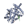
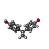
Mass: 228.286 Da / Num. of mol.: 2 / Source method: obtained synthetically / Formula: C15H16O2 / Feature type: SUBJECT OF INVESTIGATION
#4: Water ChemComp-HOH / water


Mass: 18.015 Da / Num. of mol.: 325 / Source method: isolated from a natural source / Formula: H2O
Has ligand of interestY

-
Experimental details

-
Experiment

ExperimentMethod: X-RAY DIFFRACTION / Number of used crystals: 1

-
Sample preparation

CrystalDensity Matthews: 2.34 Å3/Da / Density % sol: 47.38 %
Crystal growTemperature: 292.15 K / Method: vapor diffusion, hanging drop / pH: 7.4
Details: 20% w/v PEG 3350, 0.2 M magnesium acetate, 137 mM sodium chloride, 8.1 mM disodium hydrogen phosphate, 1.5 mM potassium dihydrogen phosphate, 2.7 mM potassium chloride, 1 mM peptide, 15 mM BPB, 1.5 % DMSO

-
Data collection

DiffractionMean temperature: 100 K / Serial crystal experiment: N
Diffraction sourceSource: SYNCHROTRON / Site: PETRA III, EMBL c/o DESY / Beamline: P14 (MX2) / Wavelength: 0.97625 Å
DetectorType: DECTRIS EIGER X 16M / Detector: PIXEL / Date: Sep 30, 2022
RadiationProtocol: SINGLE WAVELENGTH / Monochromatic (M) / Laue (L): M / Scattering type: x-ray
Radiation wavelengthWavelength: 0.97625 Å / Relative weight: 1
ReflectionResolution: 1.25→41.88 Å / Num. obs: 118830 / % possible obs: 98.9 % / Redundancy: 6.9 % / Biso Wilson estimate: 16.96 Å2 / CC1/2: 0.99 / Net I/σ(I): 12.8
Reflection shellResolution: 1.25→1.36 Å / Num. unique obs: 3027 / CC1/2: 0.615 / % possible all: 99

-
Processing

Software
NameVersionClassification
STARANISOdata scaling
PHENIX1.20.1_4487refinement
XDSdata reduction
PDB_EXTRACTdata extraction
Cootmodel building
REFMACphasing
RefinementMethod to determine structure: FOURIER SYNTHESIS / Resolution: 1.25→41.88 Å / SU ML: 0.1249 / Cross valid method: FREE R-VALUE / σ(F): 1.34 / Phase error: 23.9806
Stereochemistry target values: GeoStd + Monomer Library + CDL v1.2
RfactorNum. reflection% reflection
Rfree0.1889 5612 4.72 %
Rwork0.1622 113218 -
obs0.1634 118830 74.03 %
Solvent computationShrinkage radii: 0.9 Å / VDW probe radii: 1.1 Å / Solvent model: FLAT BULK SOLVENT MODEL
Displacement parametersBiso mean: 33.77 Å2
Refinement stepCycle: LAST / Resolution: 1.25→41.88 Å
ProteinNucleic acidLigandSolventTotal
Num. atoms2265 0 34 325 2624
Refine LS restraints
Refine-IDTypeDev idealNumber
X-RAY DIFFRACTIONf_bond_d0.00792391
X-RAY DIFFRACTIONf_angle_d0.90473241
X-RAY DIFFRACTIONf_chiral_restr0.0623375
X-RAY DIFFRACTIONf_plane_restr0.0081438
X-RAY DIFFRACTIONf_dihedral_angle_d13.9638949
LS refinement shell
Resolution (Å)Rfactor RfreeNum. reflection RfreeRfactor RworkNum. reflection RworkRefine-ID% reflection obs (%)
1.25-1.270.4057160.2613182X-RAY DIFFRACTION3.66
1.27-1.280.2517180.2812346X-RAY DIFFRACTION6.83
1.28-1.30.2744180.2788556X-RAY DIFFRACTION10.87
1.3-1.310.253280.3084738X-RAY DIFFRACTION14.46
1.31-1.330.3047580.27121006X-RAY DIFFRACTION19.9
1.33-1.350.2467680.28391297X-RAY DIFFRACTION25.92
1.35-1.370.2461960.27971760X-RAY DIFFRACTION34.35
1.37-1.390.29481280.26472297X-RAY DIFFRACTION45.58
1.39-1.410.25981250.27952862X-RAY DIFFRACTION55.25
1.41-1.430.29621640.29093506X-RAY DIFFRACTION69.85
1.43-1.460.28791970.2724011X-RAY DIFFRACTION78.33
1.46-1.490.25471900.25114462X-RAY DIFFRACTION87.1
1.49-1.510.26362510.23094805X-RAY DIFFRACTION92.58
1.51-1.550.23792830.22044865X-RAY DIFFRACTION97.3
1.55-1.580.22832880.21244975X-RAY DIFFRACTION98.06
1.58-1.620.2392550.19985031X-RAY DIFFRACTION98.88
1.62-1.660.21472460.19184979X-RAY DIFFRACTION98.42
1.66-1.70.19342260.19165070X-RAY DIFFRACTION98.42
1.7-1.750.19452730.18995008X-RAY DIFFRACTION97.98
1.75-1.810.22152470.18524963X-RAY DIFFRACTION98.92
1.81-1.870.20892880.17925045X-RAY DIFFRACTION98.89
1.87-1.950.19412460.17045054X-RAY DIFFRACTION99.05
1.95-2.040.17742360.15314989X-RAY DIFFRACTION98.88
2.04-2.140.17932290.14735058X-RAY DIFFRACTION98.14
2.14-2.280.17432330.13975040X-RAY DIFFRACTION99
2.28-2.450.15022420.13155086X-RAY DIFFRACTION99.02
2.45-2.70.16492300.13955048X-RAY DIFFRACTION98.88
2.7-3.090.16652550.15325021X-RAY DIFFRACTION98.62
3.09-3.890.18392450.14065085X-RAY DIFFRACTION99.29
3.89-41.880.17952330.15315073X-RAY DIFFRACTION99.4
Refinement TLS params.Method: refined / Origin x: 46.3993467444 Å / Origin y: 10.0579499634 Å / Origin z: 18.5375479032 Å
111213212223313233
T0.133440929889 Å20.00658539242419 Å2-0.00182808126711 Å2-0.133478707037 Å2-0.00464547578403 Å2--0.120943523064 Å2
L0.516012710088 °20.125419689536 °2-0.114541513322 °2-0.666926582282 °20.0212777573315 °2--0.490360861149 °2
S-0.00283468493823 Å °0.0768651850553 Å °-0.0230013144509 Å °-0.136738671324 Å °0.00987756691422 Å °-0.0363272656531 Å °0.0328957828525 Å °0.0186892640984 Å °-1.04898277619E-5 Å °
Refinement TLS groupSelection details: all

+
About Yorodumi

-
News

-
Feb 9, 2022. New format data for meta-information of EMDB entries

New format data for meta-information of EMDB entries

  • Version 3 of the EMDB header file is now the official format.
  • The previous official version 1.9 will be removed from the archive.

Related info.:EMDB header

External links:wwPDB to switch to version 3 of the EMDB data model

-
Aug 12, 2020. Covid-19 info

Covid-19 info

URL: https://pdbjlvh1.pdbj.org/emnavi/covid19.php

New page: Covid-19 featured information page in EM Navigator.

Related info.:Covid-19 info / Mar 5, 2020. Novel coronavirus structure data

+
Mar 5, 2020. Novel coronavirus structure data

Novel coronavirus structure data

Related info.:Yorodumi Speices / Aug 12, 2020. Covid-19 info

External links:COVID-19 featured content - PDBj / Molecule of the Month (242):Coronavirus Proteases

+
Jan 31, 2019. EMDB accession codes are about to change! (news from PDBe EMDB page)

EMDB accession codes are about to change! (news from PDBe EMDB page)

  • The allocation of 4 digits for EMDB accession codes will soon come to an end. Whilst these codes will remain in use, new EMDB accession codes will include an additional digit and will expand incrementally as the available range of codes is exhausted. The current 4-digit format prefixed with “EMD-” (i.e. EMD-XXXX) will advance to a 5-digit format (i.e. EMD-XXXXX), and so on. It is currently estimated that the 4-digit codes will be depleted around Spring 2019, at which point the 5-digit format will come into force.
  • The EM Navigator/Yorodumi systems omit the EMD- prefix.

Related info.:Q: What is EMD? / ID/Accession-code notation in Yorodumi/EM Navigator

External links:EMDB Accession Codes are Changing Soon! / Contact to PDBj

+
Jul 12, 2017. Major update of PDB

Major update of PDB

  • wwPDB released updated PDB data conforming to the new PDBx/mmCIF dictionary.
  • This is a major update changing the version number from 4 to 5, and with Remediation, in which all the entries are updated.
  • In this update, many items about electron microscopy experimental information are reorganized (e.g. em_software).
  • Now, EM Navigator and Yorodumi are based on the updated data.

External links:wwPDB Remediation / Enriched Model Files Conforming to OneDep Data Standards Now Available in the PDB FTP Archive

-
Yorodumi

Thousand views of thousand structures

  • Yorodumi is a browser for structure data from EMDB, PDB, SASBDB, etc.
  • This page is also the successor to EM Navigator detail page, and also detail information page/front-end page for Omokage search.
  • The word "yorodu" (or yorozu) is an old Japanese word meaning "ten thousand". "mi" (miru) is to see.

Related info.:EMDB / PDB / SASBDB / Comparison of 3 databanks / Yorodumi Search / Aug 31, 2016. New EM Navigator & Yorodumi / Yorodumi Papers / Jmol/JSmol / Function and homology information / Changes in new EM Navigator and Yorodumi

Read more