 Movie
Movie Controller

[English] 日本語
 Yorodumi
- PDB-9f34: Cryo-EM structure of Dopamine 3 receptor:Go complex bound to bito... -
+ Open data

- Basic information
| Entry | Database: PDB / ID: 9f34 | ||||||
|---|---|---|---|---|---|---|---|
| Title | Cryo-EM structure of Dopamine 3 receptor:Go complex bound to bitopic FOB02-04A - Conformation B | ||||||
 Components Components |
| ||||||
 Keywords Keywords | MEMBRANE PROTEIN / GPCR / complex / bitopic | ||||||
| Function / homology |  Function and homology information Function and homology informationmusculoskeletal movement, spinal reflex action / acid secretion / dopamine neurotransmitter receptor activity, coupled via Gi/Go / response to histamine / adenylate cyclase-inhibiting dopamine receptor signaling pathway / regulation of potassium ion transport / Dopamine receptors / regulation of dopamine uptake involved in synaptic transmission / positive regulation of dopamine receptor signaling pathway / phospholipase C-activating dopamine receptor signaling pathway ...musculoskeletal movement, spinal reflex action / acid secretion / dopamine neurotransmitter receptor activity, coupled via Gi/Go / response to histamine / adenylate cyclase-inhibiting dopamine receptor signaling pathway / regulation of potassium ion transport / Dopamine receptors / regulation of dopamine uptake involved in synaptic transmission / positive regulation of dopamine receptor signaling pathway / phospholipase C-activating dopamine receptor signaling pathway / negative regulation of oligodendrocyte differentiation / mu-type opioid receptor binding / : / corticotropin-releasing hormone receptor 1 binding / G-protein activation / Activation of the phototransduction cascade / Glucagon-type ligand receptors / Thromboxane signalling through TP receptor / Sensory perception of sweet, bitter, and umami (glutamate) taste / G beta:gamma signalling through PI3Kgamma / G beta:gamma signalling through CDC42 / Cooperation of PDCL (PhLP1) and TRiC/CCT in G-protein beta folding / Activation of G protein gated Potassium channels / Inhibition of voltage gated Ca2+ channels via Gbeta/gamma subunits / Ca2+ pathway / G alpha (z) signalling events / High laminar flow shear stress activates signaling by PIEZO1 and PECAM1:CDH5:KDR in endothelial cells / Glucagon-like Peptide-1 (GLP1) regulates insulin secretion / Vasopressin regulates renal water homeostasis via Aquaporins / G protein-coupled receptor internalization / Adrenaline,noradrenaline inhibits insulin secretion / ADP signalling through P2Y purinoceptor 12 / G alpha (q) signalling events / G alpha (i) signalling events / G protein-coupled dopamine receptor signaling pathway / Thrombin signalling through proteinase activated receptors (PARs) / negative regulation of synaptic transmission, glutamatergic / arachidonate secretion / photoreceptor outer segment membrane / spectrin binding / positive regulation of cytokinesis / response to morphine / negative regulation of cytosolic calcium ion concentration / alkylglycerophosphoethanolamine phosphodiesterase activity / regulation of heart contraction / dopamine metabolic process / regulation of dopamine secretion / parallel fiber to Purkinje cell synapse / negative regulation of protein secretion / photoreceptor outer segment / social behavior / postsynaptic modulation of chemical synaptic transmission / prepulse inhibition / negative regulation of blood pressure / cardiac muscle cell apoptotic process / behavioral response to cocaine / photoreceptor inner segment / positive regulation of mitotic nuclear division / adenylate cyclase-inhibiting serotonin receptor signaling pathway / G protein-coupled serotonin receptor binding / muscle contraction / bioluminescence / negative regulation of phosphatidylinositol 3-kinase/protein kinase B signal transduction / learning / circadian regulation of gene expression / response to cocaine / generation of precursor metabolites and energy / locomotory behavior / negative regulation of insulin secretion / visual learning / GABA-ergic synapse / G protein-coupled receptor activity / G-protein beta/gamma-subunit complex binding / intracellular calcium ion homeostasis / adenylate cyclase-modulating G protein-coupled receptor signaling pathway / adenylate cyclase-inhibiting G protein-coupled receptor signaling pathway / G beta:gamma signalling through PLC beta / Presynaptic function of Kainate receptors / Thromboxane signalling through TP receptor / Activation of G protein gated Potassium channels / Inhibition of voltage gated Ca2+ channels via Gbeta/gamma subunits / G-protein activation / Glucagon signaling in metabolic regulation / Prostacyclin signalling through prostacyclin receptor / G beta:gamma signalling through CDC42 / G beta:gamma signalling through BTK / ADP signalling through P2Y purinoceptor 12 / Glucagon-type ligand receptors / Adrenaline,noradrenaline inhibits insulin secretion / Vasopressin regulates renal water homeostasis via Aquaporins / Glucagon-like Peptide-1 (GLP1) regulates insulin secretion / G alpha (z) signalling events / cellular response to catecholamine stimulus / ADP signalling through P2Y purinoceptor 1 / ADORA2B mediated anti-inflammatory cytokines production / G beta:gamma signalling through PI3Kgamma / adenylate cyclase-activating dopamine receptor signaling pathway / Cooperation of PDCL (PhLP1) and TRiC/CCT in G-protein beta folding / GPER1 signaling / cellular response to prostaglandin E stimulus Similarity search - Function | ||||||
| Biological species |  Homo sapiens (human) Homo sapiens (human) | ||||||
| Method | ELECTRON MICROSCOPY / single particle reconstruction / cryo EM / Resolution: 3.09 Å | ||||||
 Authors Authors | Arroyo-Urea, S. / Garcia-Nafria, J. | ||||||
| Funding support |  Spain, 1items Spain, 1items
| ||||||
 Citation Citation | Journal: Nat Commun / Year: 2024 Title: A bitopic agonist bound to the dopamine 3 receptor reveals a selectivity site. Authors: Sandra Arroyo-Urea / Antonina L Nazarova / Ángela Carrión-Antolí / Alessandro Bonifazi / Francisco O Battiti / Jordy Homing Lam / Amy Hauck Newman / Vsevolod Katritch / Javier García-Nafría /  Abstract: Although aminergic GPCRs are the target for ~25% of approved drugs, developing subtype selective drugs is a major challenge due to the high sequence conservation at their orthosteric binding site. ...Although aminergic GPCRs are the target for ~25% of approved drugs, developing subtype selective drugs is a major challenge due to the high sequence conservation at their orthosteric binding site. Bitopic ligands are covalently joined orthosteric and allosteric pharmacophores with the potential to boost receptor selectivity and improve current medications by reducing off-target side effects. However, the lack of structural information on their binding mode impedes rational design. Here we determine the cryo-EM structure of the hDR:Gαβγ complex bound to the DR selective bitopic agonist FOB02-04A. Structural, functional and computational analyses provide insights into its binding mode and point to a new TM2-ECL1-TM1 region, which requires the N-terminal ordering of TM1, as a major determinant of subtype selectivity in aminergic GPCRs. This region is underexploited in drug development, expands the established secondary binding pocket in aminergic GPCRs and could potentially be used to design novel and subtype selective drugs. | ||||||
| History |
|
- Structure visualization
| Structure viewer | Molecule: MolmilJmol/JSmol |
|---|
- Downloads & links
-Download
| PDBx/mmCIF format | 9f34.cif.gz | 221.2 KB | Display | PDBx/mmCIF format |
|---|---|---|---|---|
| PDB format | pdb9f34.ent.gz | Display | PDB format | |
| PDBx/mmJSON format | 9f34.json.gz | Tree view | PDBx/mmJSON format | |
| Others |  Other downloads Other downloads |
-Validation report
| Arichive directory | https://data.pdbj.org/pub/pdb/validation_reports/f3/9f34ftp://data.pdbj.org/pub/pdb/validation_reports/f3/9f34 | HTTPS FTP |
|---|
-Related structure data
| Related structure data |  50169MC  9f33C M: map data used to model this data C: citing same article ( |
|---|---|
| Similar structure data |
-Links
 PDBj
PDBj


- Assembly
| Deposited unit | 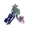
|
|---|---|
| 1 |
|
-Components
-Guanine nucleotide-binding protein ... , 3 types, 3 molecules ABC
| #1: Protein | Mass: 40097.500 Da / Num. of mol.: 1 / Mutation: S47N,G204A,E246A,M249K,A326S Source method: isolated from a genetically manipulated source Source: (gene. exp.) Homo sapiens (human) / Gene: GNAO1 / Production host:  Trichoplusia ni (cabbage looper) / References: UniProt: P09471 Trichoplusia ni (cabbage looper) / References: UniProt: P09471 |
|---|---|
| #2: Protein | Mass: 39373.992 Da / Num. of mol.: 1 Source method: isolated from a genetically manipulated source Source: (gene. exp.) Trichoplusia ni (cabbage looper) / References: UniProt: P54311 |
| #3: Protein | Mass: 7845.078 Da / Num. of mol.: 1 / Mutation: C68S Source method: isolated from a genetically manipulated source Source: (gene. exp.) Homo sapiens (human) / Gene: GNG2 / Production host: Trichoplusia ni (cabbage looper) / References: UniProt: P59768 |
-Antibody / Protein / Non-polymers , 3 types, 3 molecules ER
| #4: Antibody | Mass: 27625.844 Da / Num. of mol.: 1 Source method: isolated from a genetically manipulated source Source: (gene. exp.) Trichoplusia ni (cabbage looper) |
|---|---|
| #5: Protein | Mass: 74733.945 Da / Num. of mol.: 1 / Mutation: L119W Source method: isolated from a genetically manipulated source Details: HASS-FLAG-eGFP-D3R,HASS-FLAG-eGFP-D3R,HASS-FLAG-eGFP-D3R,HASS-FLAG-eGFP-D3R,HASS-FLAG-eGFP-D3R,HASS-FLAG-eGFP-D3R,HASS-FLAG-eGFP-D3R,HASS-FLAG-eGFP-D3R,HASS-FLAG-eGFP-D3R,HASS-FLAG-eGFP- ...Details: HASS-FLAG-eGFP-D3R,HASS-FLAG-eGFP-D3R,HASS-FLAG-eGFP-D3R,HASS-FLAG-eGFP-D3R,HASS-FLAG-eGFP-D3R,HASS-FLAG-eGFP-D3R,HASS-FLAG-eGFP-D3R,HASS-FLAG-eGFP-D3R,HASS-FLAG-eGFP-D3R,HASS-FLAG-eGFP-D3R,HASS-FLAG-eGFP-D3R,HASS-FLAG-eGFP-D3R,HASS-FLAG-eGFP-D3R,HASS-FLAG-eGFP-D3R,HASS-FLAG-eGFP-D3R,HASS-FLAG-eGFP-D3R,HASS-FLAG-eGFP-D3R,HASS-FLAG-eGFP-D3R,HASS-FLAG-eGFP-D3R,HASS-FLAG-eGFP-D3R,HASS-FLAG-eGFP-D3R,HASS-FLAG-eGFP-D3R,HASS-FLAG-eGFP-D3R,HASS-FLAG-eGFP-D3R,HASS-FLAG-eGFP-D3R,HASS-FLAG-eGFP-D3R,HASS-FLAG-eGFP-D3R,HASS-FLAG-eGFP-D3R,HASS-FLAG-eGFP-D3R,HASS-FLAG-eGFP-D3R,HASS-FLAG-eGFP-D3R,HASS-FLAG-eGFP-D3R,HASS-FLAG-eGFP-D3R,HASS-FLAG-eGFP-D3R,HASS-FLAG-eGFP-D3R,HASS-FLAG-eGFP-D3R,HASS-FLAG-eGFP-D3R,HASS-FLAG-eGFP-D3R,HASS-FLAG-eGFP-D3R,HASS-FLAG-eGFP-D3R,HASS-FLAG-eGFP-D3R,HASS-FLAG-eGFP-D3R,HASS-FLAG-eGFP-D3R,HASS-FLAG-eGFP-D3R,HASS-FLAG-eGFP-D3R,HASS-FLAG-eGFP-D3R,HASS-FLAG-eGFP-D3R,HASS-FLAG-eGFP-D3R,HASS-FLAG-eGFP-D3R,HASS-FLAG-eGFP-D3R,HASS-FLAG-eGFP-D3R,HASS-FLAG-eGFP-D3R,HASS-FLAG-eGFP-D3R,HASS-FLAG-eGFP-D3R,HASS-FLAG-eGFP-D3R,HASS-FLAG-eGFP-D3R,HASS-FLAG-eGFP-D3R,HASS-FLAG-eGFP-D3R,HASS-FLAG-eGFP-D3R,HASS-FLAG-eGFP-D3R,HASS-FLAG-eGFP-D3R,HASS-FLAG-eGFP-D3R,HASS-FLAG-eGFP-D3R,HASS-FLAG-eGFP-D3R Source: (gene. exp.) Homo sapiens (human) / Gene: GFP, DRD3 / Production host: Trichoplusia ni (cabbage looper) / References: UniProt: P42212, UniProt: P35462 |
| #6: Chemical | ChemComp-A1H9N / Mass: 433.546 Da / Num. of mol.: 1 / Source method: obtained synthetically / Formula: C25H31N5O2 / Feature type: SUBJECT OF INVESTIGATION |
-Details
| Has ligand of interest | Y |
|---|---|
| Has protein modification | Y |
-Experimental details
-Experiment
| Experiment | Method: ELECTRON MICROSCOPY |
|---|---|
| EM experiment | Aggregation state: PARTICLE / 3D reconstruction method: single particle reconstruction |
- Sample preparation
| Component | Name: D3R: Gtrimer: scFv16 complex bound to the bitopic FOB02-04A - Conformation B Type: COMPLEX / Entity ID: #1-#5 / Source: RECOMBINANT |
|---|---|
| Molecular weight | Value: 0.19 MDa / Experimental value: NO |
| Source (natural) | Organism: Homo sapiens (human) |
| Source (recombinant) | Organism: Trichoplusia ni (cabbage looper) |
| Buffer solution | pH: 7.4 |
| Specimen | Conc.: 2.8 mg/ml / Embedding applied: NO / Shadowing applied: NO / Staining applied: NO / Vitrification applied: YES |
| Specimen support | Grid material: GOLD / Grid mesh size: 300 divisions/in. / Grid type: Quantifoil R0.6/1 |
| Vitrification | Instrument: FEI VITROBOT MARK IV / Cryogen name: ETHANE / Humidity: 100 % / Chamber temperature: 277 K |
- Electron microscopy imaging
| Experimental equipment |  Model: Titan Krios / Image courtesy: FEI Company |
|---|---|
| Microscopy | Model: FEI TITAN KRIOS |
| Electron gun | Electron source:  FIELD EMISSION GUN / Accelerating voltage: 300 kV / Illumination mode: FLOOD BEAM FIELD EMISSION GUN / Accelerating voltage: 300 kV / Illumination mode: FLOOD BEAM |
| Electron lens | Mode: BRIGHT FIELD / Nominal defocus max: 3000 nm / Nominal defocus min: 1000 nm / Cs: 2.7 mm / C2 aperture diameter: 50 µm / Alignment procedure: COMA FREE |
| Specimen holder | Cryogen: NITROGEN / Specimen holder model: FEI TITAN KRIOS AUTOGRID HOLDER |
| Image recording | Electron dose: 50 e/Å2 / Detector mode: SUPER-RESOLUTION / Film or detector model: GATAN K3 (6k x 4k) / Num. of real images: 22655 |
| Image scans | Movie frames/image: 50 |
- Processing
| EM software |
| ||||||||||||||||||||||||||||||||
|---|---|---|---|---|---|---|---|---|---|---|---|---|---|---|---|---|---|---|---|---|---|---|---|---|---|---|---|---|---|---|---|---|---|
| CTF correction | Type: PHASE FLIPPING AND AMPLITUDE CORRECTION | ||||||||||||||||||||||||||||||||
| Particle selection | Num. of particles selected: 19414230 | ||||||||||||||||||||||||||||||||
| 3D reconstruction | Resolution: 3.09 Å / Resolution method: FSC 0.143 CUT-OFF / Num. of particles: 159184 / Symmetry type: POINT | ||||||||||||||||||||||||||||||||
| Atomic model building | Protocol: OTHER / Space: REAL | ||||||||||||||||||||||||||||||||
| Refine LS restraints |
|
