[English] 日本語
Yorodumi
- PDB-9f28: Crystal structure of the heterodimeric primase from pyrococcus ab... -

+
Open data


ID or keywords:

Loading...

-
Basic information

Entry
Database: PDB / ID: 9f28
Title
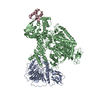
Crystal structure of the heterodimeric primase from pyrococcus abyssi (deletion of the PriL-CTD domain)
Components
  • DNA primase large subunit PriL
  • DNA primase small subunit PriS
KeywordsREPLICATION / DNA primase / PriS / PriL
Function / homology
Function and homology information


primosome complex / : / DNA-directed RNA polymerase complex / Transferases; Transferring phosphorus-containing groups; Nucleotidyltransferases / 4 iron, 4 sulfur cluster binding / metal ion binding
Similarity search - Function
DNA primase large subunit PriL / DNA primase small subunit PriS / DNA primase, small subunit, eukaryotic/archaeal / DNA primase, small subunit / DNA primase small subunit / DNA primase large subunit, eukaryotic/archaeal / Eukaryotic and archaeal DNA primase, large subunit
Similarity search - Domain/homology
FORMIC ACID / DNA primase large subunit PriL / DNA primase small subunit PriS
Similarity search - Component
Biological speciesPyrococcus abyssi (archaea)
MethodX-RAY DIFFRACTION / SYNCHROTRON / MOLECULAR REPLACEMENT / Resolution: 1.85 Å
AuthorsMadru, C. / Sauguet, L.
Funding support France, 1items
OrganizationGrant numberCountry
Agence Nationale de la Recherche (ANR)ARCHAPOL France
CitationJournal: Nat Commun / Year: 2024
Title: Communication between DNA polymerases and Replication Protein A within the archaeal replisome.
Authors: Markel Martínez-Carranza / Léa Vialle / Clément Madru / Florence Cordier / Ayten Dizkirici Tekpinar / Ahmed Haouz / Pierre Legrand / Rémy A Le Meur / Patrick England / Rémi Dulermo / J ...Authors: Markel Martínez-Carranza / Léa Vialle / Clément Madru / Florence Cordier / Ayten Dizkirici Tekpinar / Ahmed Haouz / Pierre Legrand / Rémy A Le Meur / Patrick England / Rémi Dulermo / J Iñaki Guijarro / Ghislaine Henneke / Ludovic Sauguet /
Abstract: Replication Protein A (RPA) plays a pivotal role in DNA replication by coating and protecting exposed single-stranded DNA, and acting as a molecular hub that recruits additional replication factors. ...Replication Protein A (RPA) plays a pivotal role in DNA replication by coating and protecting exposed single-stranded DNA, and acting as a molecular hub that recruits additional replication factors. We demonstrate that archaeal RPA hosts a winged-helix domain (WH) that interacts with two key actors of the replisome: the DNA primase (PriSL) and the replicative DNA polymerase (PolD). Using an integrative structural biology approach, combining nuclear magnetic resonance, X-ray crystallography and cryo-electron microscopy, we unveil how RPA interacts with PriSL and PolD through two distinct surfaces of the WH domain: an evolutionarily conserved interface and a novel binding site. Finally, RPA is shown to stimulate the activity of PriSL in a WH-dependent manner. This study provides a molecular understanding of the WH-mediated regulatory activity in central replication factors such as RPA, which regulate genome maintenance in Archaea and Eukaryotes.
History
DepositionApr 22, 2024Deposition site: PDBE / Processing site: PDBE
Revision 1.0Dec 4, 2024Provider: repository / Type: Initial release
Revision 1.1Jan 22, 2025Group: Data collection / Database references / Category: citation / citation_author / diffrn
Item: _citation.country / _citation.journal_abbrev ..._citation.country / _citation.journal_abbrev / _citation.journal_id_CSD / _citation.journal_id_ISSN / _citation.journal_volume / _citation.page_first / _citation.page_last / _citation.pdbx_database_id_DOI / _citation.pdbx_database_id_PubMed / _citation.title / _citation.year / _citation_author.identifier_ORCID / _citation_author.name / _diffrn.ambient_temp

-
Structure visualization

Structure viewerMolecule:
MolmilJmol/JSmol

Downloads & links

-
Assembly

Deposited unit
A: DNA primase small subunit PriS
B: DNA primase large subunit PriL
hetero molecules


Theoretical massNumber of molelcules
Total (without water)67,4037
Polymers67,1972
Non-polymers2065
Water11,007611
1


  • Idetical with deposited unit
  • defined by author&software
  • Evidence: gel filtration
TypeNameSymmetry operationNumber
identity operation1_555x,y,z1
Buried area2550 Å2
ΔGint-26 kcal/mol
Surface area25350 Å2
MethodPISA
Unit cell
Length a, b, c (Å)116.574, 116.574, 121.084
Angle α, β, γ (deg.)90, 90, 90
Int Tables number96
Space group name H-MP43212

-
Components

-
Protein , 2 types, 2 molecules AB

#1: Protein DNA primase small subunit PriS / DNA primase 41 kDa subunit / Pabp41 / p41


Mass: 41049.980 Da / Num. of mol.: 1
Source method: isolated from a genetically manipulated source
Source: (gene. exp.) Pyrococcus abyssi (archaea) / Gene: priS, priA, PYRAB01820, PAB2236 / Production host: Escherichia coli BL21(DE3) (bacteria)
References: UniProt: Q9V292, Transferases; Transferring phosphorus-containing groups; Nucleotidyltransferases
#2: Protein DNA primase large subunit PriL / DNA primase 46 kDa subunit / Pabp46 / p46


Mass: 26147.143 Da / Num. of mol.: 1
Source method: isolated from a genetically manipulated source
Source: (gene. exp.) Pyrococcus abyssi (archaea) / Gene: priL, priB, PYRAB01830, PAB2235 / Production host: Escherichia coli BL21(DE3) (bacteria) / References: UniProt: Q9V291

-
Non-polymers , 4 types, 616 molecules

#3: Chemical ChemComp-ZN / ZINC ION


Mass: 65.409 Da / Num. of mol.: 1 / Source method: obtained synthetically / Formula: Zn
#4: Chemical ChemComp-MG / MAGNESIUM ION


Mass: 24.305 Da / Num. of mol.: 2 / Source method: obtained synthetically / Formula: Mg
#5: Chemical ChemComp-FMT / FORMIC ACID


Mass: 46.025 Da / Num. of mol.: 2 / Source method: obtained synthetically / Formula: CH2O2
#6: Water ChemComp-HOH / water


Mass: 18.015 Da / Num. of mol.: 611 / Source method: isolated from a natural source / Formula: H2O

-
Details

Has ligand of interestN
Has protein modificationN

-
Experimental details

-
Experiment

ExperimentMethod: X-RAY DIFFRACTION / Number of used crystals: 1

-
Sample preparation

CrystalDensity Matthews: 3.06 Å3/Da / Density % sol: 59.82 %
Crystal growTemperature: 291 K / Method: vapor diffusion, hanging drop / Details: 0.2M Magnesium Formate

-
Data collection

DiffractionMean temperature: 100 K / Serial crystal experiment: N
Diffraction sourceSource: SYNCHROTRON / Site: SOLEIL / Beamline: PROXIMA 2 / Wavelength: 0.98011 Å
DetectorType: DECTRIS PILATUS 6M / Detector: PIXEL / Date: Mar 24, 2019
RadiationProtocol: SINGLE WAVELENGTH / Monochromatic (M) / Laue (L): M / Scattering type: x-ray
Radiation wavelengthWavelength: 0.98011 Å / Relative weight: 1
ReflectionResolution: 1.85→39.51 Å / Num. obs: 67117 / % possible obs: 100 % / Redundancy: 26.9 % / CC1/2: 0.999 / Rmerge(I) obs: 0.114 / Rpim(I) all: 0.023 / Net I/σ(I): 16.4
Reflection shellResolution: 1.85→1.89 Å / Rmerge(I) obs: 2.08 / Mean I/σ(I) obs: 1.6 / Num. unique obs: 4200 / CC1/2: 0.592 / Rpim(I) all: 0.609

-
Processing

Software
NameVersionClassification
BUSTER2.10.4refinement
XDSdata reduction
Aimlessdata scaling
AutoSolphasing
RefinementMethod to determine structure: MOLECULAR REPLACEMENT / Resolution: 1.85→39.51 Å / Cor.coef. Fo:Fc: 0.953 / Cor.coef. Fo:Fc free: 0.949 / SU R Cruickshank DPI: 0.126 / Cross valid method: THROUGHOUT / SU R Blow DPI: 0.136 / SU Rfree Blow DPI: 0.123 / SU Rfree Cruickshank DPI: 0.118
RfactorNum. reflection% reflectionSelection details
Rfree0.2236 3348 -RANDOM
Rwork0.1987 ---
obs0.1999 67117 93.4 %-
Displacement parametersBiso mean: 40.81 Å2
Baniso -1Baniso -2Baniso -3
1-0.6295 Å20 Å20 Å2
2--0.6295 Å20 Å2
3----1.2589 Å2
Refine analyzeLuzzati coordinate error obs: 0.23 Å
Refinement stepCycle: LAST / Resolution: 1.85→39.51 Å
ProteinNucleic acidLigandSolventTotal
Num. atoms4606 0 9 611 5226
Refine LS restraints
Refine-IDTypeDev idealNumberRestraint functionWeight
X-RAY DIFFRACTIONt_bond_d0.0094716HARMONIC2
X-RAY DIFFRACTIONt_angle_deg0.896338HARMONIC2
X-RAY DIFFRACTIONt_dihedral_angle_d1739SINUSOIDAL2
X-RAY DIFFRACTIONt_gen_planes798HARMONIC5
X-RAY DIFFRACTIONt_it4716HARMONIC10
X-RAY DIFFRACTIONt_chiral_improper_torsion580SEMIHARMONIC5
X-RAY DIFFRACTIONt_utility_distance4HARMONIC1
X-RAY DIFFRACTIONt_ideal_dist_contact4397SEMIHARMONIC4
X-RAY DIFFRACTIONt_omega_torsion3.3
X-RAY DIFFRACTIONt_other_torsion17.4
LS refinement shellResolution: 1.85→1.89 Å
RfactorNum. reflection% reflection
Rfree0.2494 59 -
Rwork0.2557 --
obs0.2554 1343 26.87 %
Refinement TLS params.

Refine-ID: X-RAY DIFFRACTION

IDL112)L122)L132)L222)L232)L332)S11 (Å °)S12 (Å °)S13 (Å °)S21 (Å °)S22 (Å °)S23 (Å °)S31 (Å °)S32 (Å °)S33 (Å °)T112)T122)T132)T222)T232)T332)Origin x (Å)Origin y (Å)Origin z (Å)
10.462-0.6618-0.01380.72510.60520.91820.12650.05630.15080.0563-0.2096-0.32610.1508-0.32610.0832-0.029-0.0677-0.05430.08070.0068-0.03063.9792-37.233528.5735
20.67460.20071.38360.65360.61652.55680.25610.20320.07340.2032-0.0588-0.45480.0734-0.4548-0.19730.0414-0.0244-0.03230.0363-0.0133-0.041913.6857-9.890259.3333
Refinement TLS group
IDRefine-IDRefine TLS-IDSelection detailsAuth asym-IDAuth seq-ID
1X-RAY DIFFRACTION1{ A|* }A1 - 344
2X-RAY DIFFRACTION1{ A|* }A350 - 352
3X-RAY DIFFRACTION2{ B|* }B1 - 211

+
About Yorodumi

-
News

-
Feb 9, 2022. New format data for meta-information of EMDB entries

New format data for meta-information of EMDB entries

  • Version 3 of the EMDB header file is now the official format.
  • The previous official version 1.9 will be removed from the archive.

Related info.:EMDB header

External links:wwPDB to switch to version 3 of the EMDB data model

-
Aug 12, 2020. Covid-19 info

Covid-19 info

URL: https://pdbj.org/emnavi/covid19.php

New page: Covid-19 featured information page in EM Navigator.

Related info.:Covid-19 info / Mar 5, 2020. Novel coronavirus structure data

+
Mar 5, 2020. Novel coronavirus structure data

Novel coronavirus structure data

Related info.:Yorodumi Speices / Aug 12, 2020. Covid-19 info

External links:COVID-19 featured content - PDBj / Molecule of the Month (242):Coronavirus Proteases

+
Jan 31, 2019. EMDB accession codes are about to change! (news from PDBe EMDB page)

EMDB accession codes are about to change! (news from PDBe EMDB page)

  • The allocation of 4 digits for EMDB accession codes will soon come to an end. Whilst these codes will remain in use, new EMDB accession codes will include an additional digit and will expand incrementally as the available range of codes is exhausted. The current 4-digit format prefixed with “EMD-” (i.e. EMD-XXXX) will advance to a 5-digit format (i.e. EMD-XXXXX), and so on. It is currently estimated that the 4-digit codes will be depleted around Spring 2019, at which point the 5-digit format will come into force.
  • The EM Navigator/Yorodumi systems omit the EMD- prefix.

Related info.:Q: What is EMD? / ID/Accession-code notation in Yorodumi/EM Navigator

External links:EMDB Accession Codes are Changing Soon! / Contact to PDBj

+
Jul 12, 2017. Major update of PDB

Major update of PDB

  • wwPDB released updated PDB data conforming to the new PDBx/mmCIF dictionary.
  • This is a major update changing the version number from 4 to 5, and with Remediation, in which all the entries are updated.
  • In this update, many items about electron microscopy experimental information are reorganized (e.g. em_software).
  • Now, EM Navigator and Yorodumi are based on the updated data.

External links:wwPDB Remediation / Enriched Model Files Conforming to OneDep Data Standards Now Available in the PDB FTP Archive

-
Yorodumi

Thousand views of thousand structures

  • Yorodumi is a browser for structure data from EMDB, PDB, SASBDB, etc.
  • This page is also the successor to EM Navigator detail page, and also detail information page/front-end page for Omokage search.
  • The word "yorodu" (or yorozu) is an old Japanese word meaning "ten thousand". "mi" (miru) is to see.

Related info.:EMDB / PDB / SASBDB / Comparison of 3 databanks / Yorodumi Search / Aug 31, 2016. New EM Navigator & Yorodumi / Yorodumi Papers / Jmol/JSmol / Function and homology information / Changes in new EM Navigator and Yorodumi

Read more