 Movie
Movie Controller


+ Open data

- Basic information
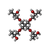
| Entry | Database: PDB / ID: 9f23 | ||||||
|---|---|---|---|---|---|---|---|
| Title | DARPin eGFP complex DP2 (2G156) | ||||||
 Components Components |
| ||||||
 Keywords Keywords | PROTEIN BINDING / DARPin / GFP | ||||||
| Function / homology | Green fluorescent protein, GFP / Green fluorescent protein-related / Green fluorescent protein / Green fluorescent protein / bioluminescence / generation of precursor metabolites and energy / : / Chem-PXN / Green fluorescent protein Function and homology information Function and homology information | ||||||
| Biological species |   Aequorea victoria (jellyfish) Aequorea victoria (jellyfish)synthetic construct (others) | ||||||
| Method |  X-RAY DIFFRACTION / SYNCHROTRON / MOLECULAR REPLACEMENT / Resolution: 1.59 Å X-RAY DIFFRACTION / SYNCHROTRON / MOLECULAR REPLACEMENT / Resolution: 1.59 Å | ||||||
 Authors Authors | Mittl, P.R. / Hansen, S. | ||||||
| Funding support |  Switzerland, 1items Switzerland, 1items
| ||||||
 Citation Citation | Journal: Commun Biol / Year: 2025 Title: Molecular features defining the efficiency of bioPROTACs. Authors: Winkelvoss, D. / Vukovic, D. / Hanny, A.C. / Riermeier, L. / Udovcic, A. / Kolibius, J. / Honegger, A. / Mittl, P.R.E. / Michel, E. / Hansen, S. / Pluckthun, A. | ||||||
| History |
|
- Structure visualization
| Structure viewer | Molecule: MolmilJmol/JSmol |
|---|
- Downloads & links
-Download
| PDBx/mmCIF format | 9f23.cif.gz | 324.1 KB | Display | PDBx/mmCIF format |
|---|---|---|---|---|
| PDB format | pdb9f23.ent.gz | Display | PDB format | |
| PDBx/mmJSON format | 9f23.json.gz | Tree view | PDBx/mmJSON format | |
| Others |  Other downloads Other downloads |
-Validation report
| Arichive directory | https://data.pdbj.org/pub/pdb/validation_reports/f2/9f23ftp://data.pdbj.org/pub/pdb/validation_reports/f2/9f23 | HTTPS FTP |
|---|
-Related structure data


-Links
 PDBj
PDBj

- Assembly
| Deposited unit | 
| ||||||||
|---|---|---|---|---|---|---|---|---|---|
| 1 | 
| ||||||||
| 2 | 
| ||||||||
| Unit cell |
|
-Components
-Protein , 2 types, 4 molecules ACBD
| #1: Protein | Mass: 27453.949 Da / Num. of mol.: 2 Source method: isolated from a genetically manipulated source Source: (gene. exp.) Aequorea victoria (jellyfish) / Gene: GFP / Production host:  #2: Protein | Mass: 13560.381 Da / Num. of mol.: 2 Source method: isolated from a genetically manipulated source Source: (gene. exp.) synthetic construct (others) / Production host: |
|---|
-Non-polymers , 6 types, 770 molecules 


| #3: Chemical | Mass: 310.384 Da / Num. of mol.: 2 / Source method: obtained synthetically / Formula: C14H30O7 #4: Chemical | ChemComp-EDO /  Mass: 62.068 Da / Num. of mol.: 15 / Source method: obtained synthetically / Formula: C2H6O2 Mass: 62.068 Da / Num. of mol.: 15 / Source method: obtained synthetically / Formula: C2H6O2#5: Chemical |  Mass: 22.990 Da / Num. of mol.: 2 / Source method: obtained synthetically / Formula: Na Mass: 22.990 Da / Num. of mol.: 2 / Source method: obtained synthetically / Formula: Na#6: Chemical | ChemComp-TRS /  Mass: 122.143 Da / Num. of mol.: 6 / Source method: obtained synthetically / Formula: C4H12NO3 / Comment: pH buffer*YM Mass: 122.143 Da / Num. of mol.: 6 / Source method: obtained synthetically / Formula: C4H12NO3 / Comment: pH buffer*YM#7: Chemical | ChemComp-PXN / ( |  Mass: 368.463 Da / Num. of mol.: 1 / Source method: obtained synthetically / Formula: C17H36O8 Mass: 368.463 Da / Num. of mol.: 1 / Source method: obtained synthetically / Formula: C17H36O8#8: Water | ChemComp-HOH / | Mass: 18.015 Da / Num. of mol.: 744 / Source method: isolated from a natural source / Formula: H2O |
|---|
-Details
| Has ligand of interest | N |
|---|---|
| Has protein modification | Y |
-Experimental details
-Experiment
| Experiment | Method: X-RAY DIFFRACTION / Number of used crystals: 1 |
|---|
- Sample preparation
| Crystal | Density Matthews: 2.25 Å3/Da / Density % sol: 45.22 % |
|---|---|
| Crystal grow | Temperature: 277.15 K / Method: vapor diffusion, sitting drop / Details: 50%pentaerythriol propoxylate 0.1 M Tris, pH 8.0 |
-Data collection
| Diffraction | Mean temperature: 100 K / Serial crystal experiment: N |
|---|---|
| Diffraction source | Source: SYNCHROTRON / Site: SLS / Beamline: X06SA / Wavelength: 1 Å |
| Detector | Type: DECTRIS EIGER X 16M / Detector: PIXEL / Date: Sep 8, 2014 |
| Radiation | Protocol: SINGLE WAVELENGTH / Monochromatic (M) / Laue (L): M / Scattering type: x-ray |
| Radiation wavelength | Wavelength: 1 Å / Relative weight: 1 |
| Reflection | Resolution: 1.59→48.08 Å / Num. obs: 95818 / % possible obs: 98.63 % / Redundancy: 6.7 % / CC1/2: 0.999 / Rmerge(I) obs: 0.086 / Rrim(I) all: 0.093 / Net I/σ(I): 14.1 |
| Reflection shell | Resolution: 1.59→1.63 Å / Num. unique obs: 3412 / CC1/2: 0.836 |
- Processing
| Software |
| ||||||||||||||||||||||||||||||||||||||||||||||||||||||||||||||||||||||||||||||||||||||||||||||||||||||||||||||||||||||||||||||||||||||||||||||||||||||||||||||||||||||||||||||||||||||
|---|---|---|---|---|---|---|---|---|---|---|---|---|---|---|---|---|---|---|---|---|---|---|---|---|---|---|---|---|---|---|---|---|---|---|---|---|---|---|---|---|---|---|---|---|---|---|---|---|---|---|---|---|---|---|---|---|---|---|---|---|---|---|---|---|---|---|---|---|---|---|---|---|---|---|---|---|---|---|---|---|---|---|---|---|---|---|---|---|---|---|---|---|---|---|---|---|---|---|---|---|---|---|---|---|---|---|---|---|---|---|---|---|---|---|---|---|---|---|---|---|---|---|---|---|---|---|---|---|---|---|---|---|---|---|---|---|---|---|---|---|---|---|---|---|---|---|---|---|---|---|---|---|---|---|---|---|---|---|---|---|---|---|---|---|---|---|---|---|---|---|---|---|---|---|---|---|---|---|---|---|---|---|---|
| Refinement | Method to determine structure: MOLECULAR REPLACEMENT / Resolution: 1.59→48.08 Å / Cor.coef. Fo:Fc: 0.975 / Cor.coef. Fo:Fc free: 0.963 / SU B: 4.669 / SU ML: 0.079 / Cross valid method: THROUGHOUT / ESU R: 0.092 / ESU R Free: 0.094 / Stereochemistry target values: MAXIMUM LIKELIHOOD / Details: HYDROGENS HAVE BEEN ADDED IN THE RIDING POSITIONS
| ||||||||||||||||||||||||||||||||||||||||||||||||||||||||||||||||||||||||||||||||||||||||||||||||||||||||||||||||||||||||||||||||||||||||||||||||||||||||||||||||||||||||||||||||||||||
| Solvent computation | Ion probe radii: 0.8 Å / Shrinkage radii: 0.8 Å / VDW probe radii: 1.2 Å / Solvent model: MASK | ||||||||||||||||||||||||||||||||||||||||||||||||||||||||||||||||||||||||||||||||||||||||||||||||||||||||||||||||||||||||||||||||||||||||||||||||||||||||||||||||||||||||||||||||||||||
| Displacement parameters | Biso mean: 17.534 Å2
| ||||||||||||||||||||||||||||||||||||||||||||||||||||||||||||||||||||||||||||||||||||||||||||||||||||||||||||||||||||||||||||||||||||||||||||||||||||||||||||||||||||||||||||||||||||||
| Refinement step | Cycle: 1 / Resolution: 1.59→48.08 Å
| ||||||||||||||||||||||||||||||||||||||||||||||||||||||||||||||||||||||||||||||||||||||||||||||||||||||||||||||||||||||||||||||||||||||||||||||||||||||||||||||||||||||||||||||||||||||
| Refine LS restraints |
|