 Movie
Movie Controller


+ Open data

- Basic information
| Entry | Database: PDB / ID: 9ew6 | ||||||
|---|---|---|---|---|---|---|---|
| Title | Binary structure of 14-3-3s and BRAF phosphopeptide (pS365) | ||||||
 Components Components |
| ||||||
 Keywords Keywords | PEPTIDE BINDING PROTEIN / 14-3-3 protein / protein-protein interaction | ||||||
| Function / homology |  Function and homology information Function and homology informationpositive regulation of axon regeneration / CD4-positive, alpha-beta T cell differentiation / CD4-positive or CD8-positive, alpha-beta T cell lineage commitment / negative regulation of synaptic vesicle exocytosis / Signalling to p38 via RIT and RIN / head morphogenesis / ARMS-mediated activation / endothelial cell apoptotic process / myeloid progenitor cell differentiation / SHOC2 M1731 mutant abolishes MRAS complex function ...positive regulation of axon regeneration / CD4-positive, alpha-beta T cell differentiation / CD4-positive or CD8-positive, alpha-beta T cell lineage commitment / negative regulation of synaptic vesicle exocytosis / Signalling to p38 via RIT and RIN / head morphogenesis / ARMS-mediated activation / endothelial cell apoptotic process / myeloid progenitor cell differentiation / SHOC2 M1731 mutant abolishes MRAS complex function / Gain-of-function MRAS complexes activate RAF signaling / negative regulation of fibroblast migration / positive regulation of D-glucose transmembrane transport / establishment of protein localization to membrane / positive regulation of axonogenesis / regulation of T cell differentiation / Negative feedback regulation of MAPK pathway / Frs2-mediated activation / stress fiber assembly / regulation of epidermal cell division / protein kinase C inhibitor activity / positive regulation of epidermal cell differentiation / keratinocyte development / keratinization / face development / regulation of cell-cell adhesion / MAP kinase kinase activity / thyroid gland development / cAMP/PKA signal transduction / Regulation of localization of FOXO transcription factors / keratinocyte proliferation / synaptic vesicle exocytosis / somatic stem cell population maintenance / positive regulation of peptidyl-serine phosphorylation / Activation of BAD and translocation to mitochondria / phosphoserine residue binding / MAP kinase kinase kinase activity / negative regulation of keratinocyte proliferation / establishment of skin barrier / negative regulation of protein localization to plasma membrane / negative regulation of endothelial cell apoptotic process / Chk1/Chk2(Cds1) mediated inactivation of Cyclin B:Cdk1 complex / SARS-CoV-2 targets host intracellular signalling and regulatory pathways / postsynaptic modulation of chemical synaptic transmission / negative regulation of protein kinase activity / negative regulation of stem cell proliferation / RHO GTPases activate PKNs / SARS-CoV-1 targets host intracellular signalling and regulatory pathways / positive regulation of protein localization / positive regulation of stress fiber assembly / ERK1 and ERK2 cascade / positive regulation of substrate adhesion-dependent cell spreading / substrate adhesion-dependent cell spreading / positive regulation of cell adhesion / protein sequestering activity / negative regulation of innate immune response / cellular response to calcium ion / TP53 Regulates Transcription of Genes Involved in G2 Cell Cycle Arrest / protein export from nucleus / release of cytochrome c from mitochondria / thymus development / animal organ morphogenesis / stem cell proliferation / positive regulation of protein export from nucleus / TP53 Regulates Metabolic Genes / Translocation of SLC2A4 (GLUT4) to the plasma membrane / RAF activation / Spry regulation of FGF signaling / Signaling by high-kinase activity BRAF mutants / MAP2K and MAPK activation / visual learning / cellular response to xenobiotic stimulus / epidermal growth factor receptor signaling pathway / centriolar satellite / long-term synaptic potentiation / intrinsic apoptotic signaling pathway in response to DNA damage / Negative regulation of MAPK pathway / Signaling by RAF1 mutants / Signaling by moderate kinase activity BRAF mutants / Paradoxical activation of RAF signaling by kinase inactive BRAF / Signaling downstream of RAS mutants / Signaling by BRAF and RAF1 fusions / intracellular protein localization / T cell differentiation in thymus / T cell receptor signaling pathway / MAPK cascade / regulation of cell population proliferation / presynapse / regulation of protein localization / cell body / positive regulation of cell growth / scaffold protein binding / negative regulation of neuron apoptotic process / protein phosphorylation / positive regulation of ERK1 and ERK2 cascade / protein kinase activity / non-specific serine/threonine protein kinase / neuron projection / postsynapse / regulation of cell cycle Similarity search - Function | ||||||
| Biological species |  Homo sapiens (human) Homo sapiens (human) | ||||||
| Method |  X-RAY DIFFRACTION / SYNCHROTRON / MOLECULAR REPLACEMENT / Resolution: 1.65 Å X-RAY DIFFRACTION / SYNCHROTRON / MOLECULAR REPLACEMENT / Resolution: 1.65 Å | ||||||
 Authors Authors | Konstantinidou, M. / Vickery, H. / Pennings, M.A.M. / Virta, J. / Visser, E.J. / Oetelaar, M.C.M. / Overmans, M. / Neitz, J. / Ottmann, C. / Brunsveld, L. / Arkin, M.R. | ||||||
| Funding support |  United States, 1items United States, 1items
| ||||||
 Citation Citation | Journal: To Be Published Title: Small molecule stabilization of the 14-3-3sigma/CRAF complex inhibits the MAPK pathway Authors: Konstantinidou, M. / Vickery, H. / Pennings, M.A.M. / Virta, J. / Visser, E.J. / Oetelaar, M.C.M. / Overmans, M. / Neitz, J. / Ottmann, C. / Brunsveld, L. / Arkin, M.R. | ||||||
| History |
|
- Structure visualization
| Structure viewer | Molecule: MolmilJmol/JSmol |
|---|
- Downloads & links
-Download
| PDBx/mmCIF format | 9ew6.cif.gz | 117.4 KB | Display | PDBx/mmCIF format |
|---|---|---|---|---|
| PDB format | pdb9ew6.ent.gz | 88.4 KB | Display | PDB format |
| PDBx/mmJSON format | 9ew6.json.gz | Tree view | PDBx/mmJSON format | |
| Others |  Other downloads Other downloads |
-Validation report
| Summary document | 9ew6_validation.pdf.gz | 433.6 KB | Display | wwPDB validaton report |
|---|---|---|---|---|
| Full document | 9ew6_full_validation.pdf.gz | 433.9 KB | Display | |
| Data in XML | 9ew6_validation.xml.gz | 14.1 KB | Display | |
| Data in CIF | 9ew6_validation.cif.gz | 19.3 KB | Display | |
| Arichive directory | https://data.pdbj.org/pub/pdb/validation_reports/ew/9ew6ftp://data.pdbj.org/pub/pdb/validation_reports/ew/9ew6 | HTTPS FTP |
-Related structure data
| Related structure data |  8q55C  8q5cC  8qs2C 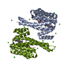 8qs3C  8qs4C  8qs5C  8qs6C  8qs7C  8qs8C  8qs9C  8qsaC  8qsbC  8qscC  8qsdC  8qseC  8qsfC  8qsgC  8qshC  8s42C  9ew1C  9ew3C 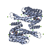 9ew4C  9ew5C  9ew7C C: citing same article ( |
|---|---|
| Similar structure data |
-Links
 PDBj
PDBj


- Assembly
| Deposited unit | 
| ||||||||
|---|---|---|---|---|---|---|---|---|---|
| 1 | 
| ||||||||
| Unit cell |
|
-Components
| #1: Protein | Mass: 26542.914 Da / Num. of mol.: 1 Source method: isolated from a genetically manipulated source Source: (gene. exp.) Homo sapiens (human) / Gene: SFN, HME1 / Production host:  | ||||||||
|---|---|---|---|---|---|---|---|---|---|
| #2: Protein/peptide | Mass: 1012.935 Da / Num. of mol.: 1 / Source method: obtained synthetically / Source: (synth.) Homo sapiens (human)References: UniProt: P15056, non-specific serine/threonine protein kinase | ||||||||
| #3: Chemical |   Mass: 35.453 Da / Num. of mol.: 2 / Source method: obtained synthetically / Formula: Cl Mass: 35.453 Da / Num. of mol.: 2 / Source method: obtained synthetically / Formula: Cl#4: Chemical | ChemComp-MG / |   Mass: 24.305 Da / Num. of mol.: 1 / Source method: obtained synthetically / Formula: Mg Mass: 24.305 Da / Num. of mol.: 1 / Source method: obtained synthetically / Formula: Mg#5: Water | ChemComp-HOH / |  Mass: 18.015 Da / Num. of mol.: 186 / Source method: isolated from a natural source / Formula: H2O Mass: 18.015 Da / Num. of mol.: 186 / Source method: isolated from a natural source / Formula: H2OHas ligand of interest | N | Has protein modification | Y | |
-Experimental details
-Experiment
| Experiment | Method: X-RAY DIFFRACTION / Number of used crystals: 1 |
|---|
- Sample preparation
| Crystal | Density Matthews: 2.97 Å3/Da / Density % sol: 58.52 % |
|---|---|
| Crystal grow | Temperature: 277 K / Method: vapor diffusion, sitting drop Details: 0.095 M HEPES pH=7.1-7.7 0.19 M CaCl2 5% glycerol 24-29% PEG400 |
-Data collection
| Diffraction | Mean temperature: 100 K / Serial crystal experiment: N |
|---|---|
| Diffraction source | Source: SYNCHROTRON / Site: ESRF  / Beamline: ID30B / Wavelength: 0.873128 Å / Beamline: ID30B / Wavelength: 0.873128 Å |
| Detector | Type: DECTRIS EIGER2 X 9M / Detector: PIXEL / Date: Feb 19, 2024 |
| Radiation | Protocol: SINGLE WAVELENGTH / Monochromatic (M) / Laue (L): M / Scattering type: x-ray |
| Radiation wavelength | Wavelength: 0.873128 Å / Relative weight: 1 |
| Reflection | Resolution: 1.65→80.44 Å / Num. obs: 38175 / % possible obs: 99.3 % / Redundancy: 13.5 % / CC1/2: 0.998 / Net I/σ(I): 14.4 |
| Reflection shell | Resolution: 1.65→1.68 Å / Mean I/σ(I) obs: 1.4 / Num. unique obs: 1908 / CC1/2: 0.694 |
- Processing
| Software |
| ||||||||||||||||||||||||||||||||||||||||||||||||||||||||||||||||||||||||||||||||||||||||||||||||||||
|---|---|---|---|---|---|---|---|---|---|---|---|---|---|---|---|---|---|---|---|---|---|---|---|---|---|---|---|---|---|---|---|---|---|---|---|---|---|---|---|---|---|---|---|---|---|---|---|---|---|---|---|---|---|---|---|---|---|---|---|---|---|---|---|---|---|---|---|---|---|---|---|---|---|---|---|---|---|---|---|---|---|---|---|---|---|---|---|---|---|---|---|---|---|---|---|---|---|---|---|---|---|
| Refinement | Method to determine structure: MOLECULAR REPLACEMENT / Resolution: 1.65→62.27 Å / Cor.coef. Fo:Fc: 0.966 / Cor.coef. Fo:Fc free: 0.958 / SU B: 6.792 / SU ML: 0.094 / Cross valid method: THROUGHOUT / ESU R: 0.107 / ESU R Free: 0.089 / Stereochemistry target values: MAXIMUM LIKELIHOOD Details: HYDROGENS HAVE BEEN USED IF PRESENT IN THE INPUT U VALUES : REFINED INDIVIDUALLY
| ||||||||||||||||||||||||||||||||||||||||||||||||||||||||||||||||||||||||||||||||||||||||||||||||||||
| Solvent computation | Ion probe radii: 0.8 Å / Shrinkage radii: 0.8 Å / VDW probe radii: 1 Å / Solvent model: MASK | ||||||||||||||||||||||||||||||||||||||||||||||||||||||||||||||||||||||||||||||||||||||||||||||||||||
| Displacement parameters | Biso mean: 37.648 Å2
| ||||||||||||||||||||||||||||||||||||||||||||||||||||||||||||||||||||||||||||||||||||||||||||||||||||
| Refinement step | Cycle: LAST / Resolution: 1.65→62.27 Å
| ||||||||||||||||||||||||||||||||||||||||||||||||||||||||||||||||||||||||||||||||||||||||||||||||||||
| Refine LS restraints |
| ||||||||||||||||||||||||||||||||||||||||||||||||||||||||||||||||||||||||||||||||||||||||||||||||||||
| LS refinement shell | Resolution: 1.65→1.693 Å / Total num. of bins used: 20
|
