 Movie
Movie Controller

[English] 日本語
 Yorodumi
- PDB-9evk: Crystal Structure of human Collagen Hydroxylysine Galactosyltrans... -
+ Open data

- Basic information
| Entry | Database: PDB / ID: 9evk | |||||||||||||||
|---|---|---|---|---|---|---|---|---|---|---|---|---|---|---|---|---|
| Title | Crystal Structure of human Collagen Hydroxylysine Galactosyltransferase GLT25D1/COLGALT1 | |||||||||||||||
 Components Components | Procollagen galactosyltransferase 1 | |||||||||||||||
 Keywords Keywords | TRANSFERASE / Collagen Biosynthesis / Extracellular Matrix / Post-translational modifications / Glycosyltransferase | |||||||||||||||
| Function / homology |  Function and homology information Function and homology informationprocollagen galactosyltransferase / procollagen galactosyltransferase activity / positive regulation of collagen fibril organization / Collagen biosynthesis and modifying enzymes / protein O-linked glycosylation / collagen fibril organization / endoplasmic reticulum lumen / membrane Similarity search - Function | |||||||||||||||
| Biological species |  Homo sapiens (human) Homo sapiens (human) | |||||||||||||||
| Method |  X-RAY DIFFRACTION / SYNCHROTRON / MOLECULAR REPLACEMENT / Resolution: 3 Å X-RAY DIFFRACTION / SYNCHROTRON / MOLECULAR REPLACEMENT / Resolution: 3 Å | |||||||||||||||
 Authors Authors | De Marco, M. / Scietti, L. / Rai, S.R. / Mattoteia, D. / Pinnola, A. / Forneris, F. | |||||||||||||||
| Funding support |  Italy, Italy,  United States, United States,  Japan, 4items Japan, 4items
| |||||||||||||||
 Citation Citation | Journal: Nat Commun / Year: 2025 Title: Molecular structure and enzymatic mechanism of the human collagen hydroxylysine galactosyltransferase GLT25D1/COLGALT1. Authors: De Marco, M. / Rai, S.R. / Scietti, L. / Mattoteia, D. / Liberi, S. / Moroni, E. / Pinnola, A. / Vetrano, A. / Iacobucci, C. / Santambrogio, C. / Colombo, G. / Forneris, F. | |||||||||||||||
| History |
|
- Structure visualization
| Structure viewer | Molecule: MolmilJmol/JSmol |
|---|
- Downloads & links
-Download
| PDBx/mmCIF format | 9evk.cif.gz | 445.1 KB | Display | PDBx/mmCIF format |
|---|---|---|---|---|
| PDB format | pdb9evk.ent.gz | 366.6 KB | Display | PDB format |
| PDBx/mmJSON format | 9evk.json.gz | Tree view | PDBx/mmJSON format | |
| Others |  Other downloads Other downloads |
-Validation report
| Arichive directory | https://data.pdbj.org/pub/pdb/validation_reports/ev/9evkftp://data.pdbj.org/pub/pdb/validation_reports/ev/9evk | HTTPS FTP |
|---|
-Related structure data
| Related structure data |  9evjC  9evlC C: citing same article ( |
|---|---|
| Similar structure data | |
| Other databases |
|
-Links
 PDBj
PDBj


- Assembly
| Deposited unit | 
| ||||||||
|---|---|---|---|---|---|---|---|---|---|
| 1 | 
| ||||||||
| Unit cell |
|
-Components
-Protein , 1 types, 2 molecules AB
| #1: Protein | Mass: 68589.164 Da / Num. of mol.: 2 Source method: isolated from a genetically manipulated source Details: N-terminal GS and C-terminal AAA are restriction scars. Residues not modeled are flexible Source: (gene. exp.) Homo sapiens (human) / Gene: COLGALT1, GLT25D1, PSEC0241 / Production host:  References: UniProt: Q8NBJ5, procollagen galactosyltransferase |
|---|
-Non-polymers , 6 types, 22 molecules 
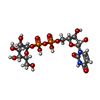


| #2: Chemical | ChemComp-CL /  Mass: 35.453 Da / Num. of mol.: 6 / Source method: obtained synthetically / Formula: Cl Mass: 35.453 Da / Num. of mol.: 6 / Source method: obtained synthetically / Formula: Cl#3: Chemical |  Mass: 566.302 Da / Num. of mol.: 2 / Source method: obtained synthetically / Formula: C15H24N2O17P2 / Feature type: SUBJECT OF INVESTIGATION Mass: 566.302 Da / Num. of mol.: 2 / Source method: obtained synthetically / Formula: C15H24N2O17P2 / Feature type: SUBJECT OF INVESTIGATION#4: Chemical |  Mass: 195.237 Da / Num. of mol.: 2 / Source method: obtained synthetically / Formula: C6H13NO4S / Comment: pH buffer*YM Mass: 195.237 Da / Num. of mol.: 2 / Source method: obtained synthetically / Formula: C6H13NO4S / Comment: pH buffer*YM#5: Chemical |  Mass: 40.078 Da / Num. of mol.: 2 / Source method: obtained synthetically / Formula: Ca / Feature type: SUBJECT OF INVESTIGATION Mass: 40.078 Da / Num. of mol.: 2 / Source method: obtained synthetically / Formula: Ca / Feature type: SUBJECT OF INVESTIGATION#6: Chemical | ChemComp-NA / |  Mass: 22.990 Da / Num. of mol.: 1 / Source method: obtained synthetically / Formula: Na Mass: 22.990 Da / Num. of mol.: 1 / Source method: obtained synthetically / Formula: Na#7: Water | ChemComp-HOH / | Mass: 18.015 Da / Num. of mol.: 9 / Source method: isolated from a natural source / Formula: H2O |
|---|
-Details
| Has ligand of interest | Y |
|---|---|
| Has protein modification | Y |
-Experimental details
-Experiment
| Experiment | Method: X-RAY DIFFRACTION / Number of used crystals: 1 |
|---|
- Sample preparation
| Crystal | Density Matthews: 3.32 Å3/Da / Density % sol: 0.62 % |
|---|---|
| Crystal grow | Temperature: 293 K / Method: vapor diffusion, hanging drop / pH: 6.5 Details: 8% poly-ethylene-glycol (PEG) MW 4000, 100 mM 2-(N-morpholino)ethanesulfonic acid (MES)/NaOH, 20% glycerol, pH 6.5 |
-Data collection
| Diffraction | Mean temperature: 100 K / Serial crystal experiment: N |
|---|---|
| Diffraction source | Source: SYNCHROTRON / Site: ESRF  / Beamline: ID23-1 / Wavelength: 0.8856 Å / Beamline: ID23-1 / Wavelength: 0.8856 Å |
| Detector | Type: DECTRIS EIGER X 16M / Detector: PIXEL / Date: Sep 25, 2022 |
| Radiation | Protocol: SINGLE WAVELENGTH / Monochromatic (M) / Laue (L): M / Scattering type: x-ray |
| Radiation wavelength | Wavelength: 0.8856 Å / Relative weight: 1 |
| Reflection | Resolution: 3→49.62 Å / Num. obs: 35735 / % possible obs: 98.4 % / Redundancy: 3.5 % / CC1/2: 0.992 / Rmerge(I) obs: 0.13 / Rpim(I) all: 0.101 / Net I/σ(I): 6.2 |
| Reflection shell | Resolution: 3→3.13 Å / Rmerge(I) obs: 1.047 / Mean I/σ(I) obs: 1 / Num. unique obs: 4385 / CC1/2: 0.402 / Rpim(I) all: 0.811 |
- Processing
| Software |
| ||||||||||||||||||||||||||||||||||||||||||||||||||||||||||||||||||||||||||||||||||||||||||||||||||||||||||||||||||||||||||||||||||||||||||||||||||||||||||||||||||||||||||||||||||||||||||||||||||||||||
|---|---|---|---|---|---|---|---|---|---|---|---|---|---|---|---|---|---|---|---|---|---|---|---|---|---|---|---|---|---|---|---|---|---|---|---|---|---|---|---|---|---|---|---|---|---|---|---|---|---|---|---|---|---|---|---|---|---|---|---|---|---|---|---|---|---|---|---|---|---|---|---|---|---|---|---|---|---|---|---|---|---|---|---|---|---|---|---|---|---|---|---|---|---|---|---|---|---|---|---|---|---|---|---|---|---|---|---|---|---|---|---|---|---|---|---|---|---|---|---|---|---|---|---|---|---|---|---|---|---|---|---|---|---|---|---|---|---|---|---|---|---|---|---|---|---|---|---|---|---|---|---|---|---|---|---|---|---|---|---|---|---|---|---|---|---|---|---|---|---|---|---|---|---|---|---|---|---|---|---|---|---|---|---|---|---|---|---|---|---|---|---|---|---|---|---|---|---|---|---|---|---|
| Refinement | Method to determine structure: MOLECULAR REPLACEMENT / Resolution: 3→49.62 Å / SU ML: 0.45 / Cross valid method: FREE R-VALUE / σ(F): 1.35 / Phase error: 27.81 / Stereochemistry target values: ML
| ||||||||||||||||||||||||||||||||||||||||||||||||||||||||||||||||||||||||||||||||||||||||||||||||||||||||||||||||||||||||||||||||||||||||||||||||||||||||||||||||||||||||||||||||||||||||||||||||||||||||
| Solvent computation | Shrinkage radii: 0.9 Å / VDW probe radii: 1.1 Å / Solvent model: FLAT BULK SOLVENT MODEL | ||||||||||||||||||||||||||||||||||||||||||||||||||||||||||||||||||||||||||||||||||||||||||||||||||||||||||||||||||||||||||||||||||||||||||||||||||||||||||||||||||||||||||||||||||||||||||||||||||||||||
| Refinement step | Cycle: LAST / Resolution: 3→49.62 Å
| ||||||||||||||||||||||||||||||||||||||||||||||||||||||||||||||||||||||||||||||||||||||||||||||||||||||||||||||||||||||||||||||||||||||||||||||||||||||||||||||||||||||||||||||||||||||||||||||||||||||||
| Refine LS restraints |
| ||||||||||||||||||||||||||||||||||||||||||||||||||||||||||||||||||||||||||||||||||||||||||||||||||||||||||||||||||||||||||||||||||||||||||||||||||||||||||||||||||||||||||||||||||||||||||||||||||||||||
| LS refinement shell |
| ||||||||||||||||||||||||||||||||||||||||||||||||||||||||||||||||||||||||||||||||||||||||||||||||||||||||||||||||||||||||||||||||||||||||||||||||||||||||||||||||||||||||||||||||||||||||||||||||||||||||
| Refinement TLS params. | Method: refined / Refine-ID: X-RAY DIFFRACTION
| ||||||||||||||||||||||||||||||||||||||||||||||||||||||||||||||||||||||||||||||||||||||||||||||||||||||||||||||||||||||||||||||||||||||||||||||||||||||||||||||||||||||||||||||||||||||||||||||||||||||||
| Refinement TLS group |
|
