| Entry | Database: PDB / ID: 9eou
|
|---|
| Title | Crystal Structure of the b1b2 domains from Human Neuropilin-1 in complex with a peptide. |
|---|
 Components Components | |
|---|
 Keywords Keywords | STRUCTURAL PROTEIN / angiogenesis / Neuropilin-1 |
|---|
| Function / homology |  Function and homology information Function and homology information
negative regulation of collateral sprouting of intact axon in response to injury / positive regulation of estradiol secretion / androgen catabolic process / endothelial tip cell fate specification / basal dendrite development / otic placode development / protein localization to early endosome / basal dendrite arborization / dichotomous subdivision of terminal units involved in salivary gland branching / retina vasculature morphogenesis in camera-type eye ...negative regulation of collateral sprouting of intact axon in response to injury / positive regulation of estradiol secretion / androgen catabolic process / endothelial tip cell fate specification / basal dendrite development / otic placode development / protein localization to early endosome / basal dendrite arborization / dichotomous subdivision of terminal units involved in salivary gland branching / retina vasculature morphogenesis in camera-type eye / vestibulocochlear nerve structural organization / dorsal root ganglion morphogenesis / ventral trunk neural crest cell migration / sympathetic neuron projection guidance / facioacoustic ganglion development / trigeminal ganglion development / biomineral tissue development / trigeminal nerve structural organization / sensory neuron axon guidance / postsynapse organization / facial nerve structural organization / branchiomotor neuron axon guidance / gonadotrophin-releasing hormone neuronal migration to the hypothalamus / negative regulation of axon extension involved in axon guidance / response to macrophage colony-stimulating factor / axon extension involved in axon guidance / VEGF-activated neuropilin signaling pathway / renal artery morphogenesis / neurofilament / sympathetic neuron projection extension / Neurophilin interactions with VEGF and VEGFR / vascular endothelial growth factor binding / regulation of vascular endothelial growth factor receptor signaling pathway / angiogenesis involved in coronary vascular morphogenesis / motor neuron migration / response to 2,3,7,8-tetrachlorodibenzodioxine / neural crest cell migration involved in autonomic nervous system development / sympathetic ganglion development / positive regulation of axon extension involved in axon guidance / axonogenesis involved in innervation / vascular endothelial growth factor receptor activity / CHL1 interactions / endothelial cell chemotaxis / cellular response to testosterone stimulus / regulation of vesicle-mediated transport / semaphorin receptor complex / neuropilin signaling pathway / Signaling by ROBO receptors / SEMA3A-Plexin repulsion signaling by inhibiting Integrin adhesion / substrate-dependent cell migration, cell extension / Signaling by PDGF / commissural neuron axon guidance / coronary artery morphogenesis / semaphorin receptor activity / RUNX3 Regulates Immune Response and Cell Migration / CRMPs in Sema3A signaling / outflow tract septum morphogenesis / positive regulation of bone resorption / motor neuron axon guidance / axonal fasciculation / hepatocyte growth factor receptor signaling pathway / cell migration involved in sprouting angiogenesis / sprouting angiogenesis / extracellular matrix binding / retinal ganglion cell axon guidance / response to steroid hormone / regulation of Cdc42 protein signal transduction / response to vitamin D / positive regulation of cell migration involved in sprouting angiogenesis / neural crest cell migration / artery morphogenesis / positive regulation of filopodium assembly / positive regulation of smooth muscle cell migration / cellular response to hepatocyte growth factor stimulus / growth factor binding / Mechanical load activates signaling by PIEZO1 and integrins in osteocytes / branching involved in blood vessel morphogenesis / positive chemotaxis / cytokine binding / sorting endosome / small molecule binding / platelet-derived growth factor receptor signaling pathway / decidualization / semaphorin-plexin signaling pathway / positive regulation of phosphorylation / Sema3A PAK dependent Axon repulsion / cellular response to vascular endothelial growth factor stimulus / positive regulation of focal adhesion assembly / vascular endothelial growth factor receptor signaling pathway / Integrin cell surface interactions / vasculogenesis / coreceptor activity / positive regulation of stress fiber assembly / Degradation of the extracellular matrix / positive regulation of endothelial cell proliferation / positive regulation of substrate adhesion-dependent cell spreading / positive regulation of endothelial cell migration / embryo implantation / axon guidance / GTPase activator activitySimilarity search - Function Osteopontin / Osteopontin, conserved site / Osteopontin / Osteopontin signature. / Osteopontin / Neuropilin / Neuropilin, C-terminal / C-terminal domain of neuropilin glycoprotein / MAM domain signature. / Domain in meprin, A5, receptor protein tyrosine phosphatase mu (and others) ...Osteopontin / Osteopontin, conserved site / Osteopontin / Osteopontin signature. / Osteopontin / Neuropilin / Neuropilin, C-terminal / C-terminal domain of neuropilin glycoprotein / MAM domain signature. / Domain in meprin, A5, receptor protein tyrosine phosphatase mu (and others) / : / Coagulation factors 5/8 type C domain (FA58C) signature 2. / MAM domain, meprin/A5/mu / MAM domain / MAM domain profile. / Coagulation factors 5/8 type C domain (FA58C) signature 1. / CUB domain / Domain first found in C1r, C1s, uEGF, and bone morphogenetic protein. / CUB domain / Spermadhesin, CUB domain superfamily / CUB domain profile. / Coagulation factor 5/8 C-terminal domain, discoidin domain / Coagulation factors 5/8 type C domain (FA58C) profile. / F5/8 type C domain / Coagulation factor 5/8 C-terminal domain / Galactose-binding-like domain superfamily / Concanavalin A-like lectin/glucanase domain superfamilySimilarity search - Domain/homology |
|---|
| Biological species |  Homo sapiens (human) Homo sapiens (human) |
|---|
| Method |  X-RAY DIFFRACTION / SYNCHROTRON / MOLECULAR REPLACEMENT / Resolution: 1.55 Å X-RAY DIFFRACTION / SYNCHROTRON / MOLECULAR REPLACEMENT / Resolution: 1.55 Å |
|---|
 Authors Authors | Caing-Carlsson, R. / Duelli, A. / Walse, B. |
|---|
| Funding support |  Denmark, Denmark,  Sweden, 3items Sweden, 3items | Organization | Grant number | Country |
|---|
| Novo Nordisk Foundation | | Denmark | | Swedish Research Council | | Sweden | | Vinnova | | Sweden |
|
|---|
 Citation Citation | Journal: Pharmacol Res / Year: 2024
Title: Identification of an osteopontin-derived peptide that binds neuropilin-1 and activates vascular repair responses and angiogenesis.
Authors: Chen, Y. / Gialeli, C. / Shen, J. / Duner, P. / Walse, B. / Duelli, A. / Caing-Carlsson, R. / Blom, A.M. / Zibert, J.R. / Nilsson, A.H. / Alenfall, J. / Liang, C. / Nilsson, J. |
|---|
| History | | Deposition | Mar 15, 2024 | Deposition site: PDBE / Processing site: PDBE |
|---|
| Revision 1.0 | Jun 26, 2024 | Provider: repository / Type: Initial release |
|---|
| Revision 1.1 | Oct 9, 2024 | Group: Structure summary / Category: pdbx_entry_details / pdbx_modification_feature |
|---|
|
|---|
 Movie
Movie Controller

 Yorodumi
 Open data
 Basic information
 Structure visualization
 Downloads & links
 Other downloads
 PDBj
PDBj


 Assembly
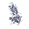

 Mass: 18.015 Da / Num. of mol.: 231 / Source method: isolated from a natural source / Formula: H2O
 Sample preparation
 / Beamline: I04 / Wavelength: 0.9795 Å
 Processing