 Movie
Movie Controller

[English] 日本語
 Yorodumi
- PDB-9eif: A broad-substrate spectrum lactate racemase A from Isosphaera pal... -
+ Open data

- Basic information
| Entry | Database: PDB / ID: 9eif | |||||||||
|---|---|---|---|---|---|---|---|---|---|---|
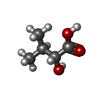
| Title | A broad-substrate spectrum lactate racemase A from Isosphaera pallida in complex with D-2-Hydroxyisovalerate | |||||||||
 Components Components | A broad-substrate spectrum lactate racemase A | |||||||||
 Keywords Keywords | ISOMERASE / Catalytic activity / isomerase activity / racemase and epimerase activity racemase acting on hydroxy acids and derivatives | |||||||||
| Function / homology |  Function and homology information Function and homology information | |||||||||
| Biological species |  Isosphaera pallida (bacteria) Isosphaera pallida (bacteria) | |||||||||
| Method |  X-RAY DIFFRACTION / SYNCHROTRON / MOLECULAR REPLACEMENT / Resolution: 1.65 Å X-RAY DIFFRACTION / SYNCHROTRON / MOLECULAR REPLACEMENT / Resolution: 1.65 Å | |||||||||
 Authors Authors | Gatreddi, S. / Hausinger, R.P. / Hu, J. | |||||||||
| Funding support |  United States, 2items United States, 2items
| |||||||||
 Citation Citation | Journal: Biorxiv / Year: 2024 Title: Structural Basis for Catalysis and Substrate Specificity of a LarA Racemase with a Broad Substrate Spectrum. Authors: Gatreddi, S. / Urdiain-Arraiza, J. / Desguin, B. / Hausinger, R.P. / Hu, J. | |||||||||
| History |
|
- Structure visualization
| Structure viewer | Molecule: MolmilJmol/JSmol |
|---|
- Downloads & links
-Download
| PDBx/mmCIF format | 9eif.cif.gz | 190.4 KB | Display | PDBx/mmCIF format |
|---|---|---|---|---|
| PDB format | pdb9eif.ent.gz | 146.8 KB | Display | PDB format |
| PDBx/mmJSON format | 9eif.json.gz | Tree view | PDBx/mmJSON format | |
| Others |  Other downloads Other downloads |
-Validation report
| Arichive directory | https://data.pdbj.org/pub/pdb/validation_reports/ei/9eifftp://data.pdbj.org/pub/pdb/validation_reports/ei/9eif | HTTPS FTP |
|---|
-Related structure data


-Links
 PDBj
PDBj- Assembly
| Deposited unit | 
| ||||||||
|---|---|---|---|---|---|---|---|---|---|
| 1 | 
| ||||||||
| 2 | 
| ||||||||
| Unit cell |
|
-Components
-Protein , 1 types, 2 molecules AB
| #1: Protein | Mass: 45898.801 Da / Num. of mol.: 2 Source method: isolated from a genetically manipulated source Source: (gene. exp.) Isosphaera pallida (bacteria) / Gene: Isop_3476 / Production host: Lactococcus lactis (lactic acid bacteria) / Strain (production host): NZ3900 / References: UniProt: E8QWZ4 |
|---|
-Non-polymers , 6 types, 644 molecules 


| #2: Chemical |  Mass: 396.353 Da / Num. of mol.: 2 / Source method: obtained synthetically / Formula: C12H15NO8PS2 / Feature type: SUBJECT OF INVESTIGATION Mass: 396.353 Da / Num. of mol.: 2 / Source method: obtained synthetically / Formula: C12H15NO8PS2 / Feature type: SUBJECT OF INVESTIGATION#3: Chemical | ChemComp-PGE / |  Mass: 150.173 Da / Num. of mol.: 1 / Source method: obtained synthetically / Formula: C6H14O4 Mass: 150.173 Da / Num. of mol.: 1 / Source method: obtained synthetically / Formula: C6H14O4#4: Chemical |  Mass: 106.120 Da / Num. of mol.: 2 / Source method: obtained synthetically / Formula: C4H10O3 Mass: 106.120 Da / Num. of mol.: 2 / Source method: obtained synthetically / Formula: C4H10O3#5: Chemical |  Type: L-peptide linking / Mass: 118.131 Da / Num. of mol.: 2 / Source method: obtained synthetically / Formula: C5H10O3 / Feature type: SUBJECT OF INVESTIGATION Type: L-peptide linking / Mass: 118.131 Da / Num. of mol.: 2 / Source method: obtained synthetically / Formula: C5H10O3 / Feature type: SUBJECT OF INVESTIGATION#6: Chemical |  Mass: 58.693 Da / Num. of mol.: 2 / Source method: obtained synthetically / Formula: Ni / Feature type: SUBJECT OF INVESTIGATION Mass: 58.693 Da / Num. of mol.: 2 / Source method: obtained synthetically / Formula: Ni / Feature type: SUBJECT OF INVESTIGATION#7: Water | ChemComp-HOH / | Mass: 18.015 Da / Num. of mol.: 635 / Source method: isolated from a natural source / Formula: H2O |
|---|
-Details
| Has ligand of interest | Y |
|---|---|
| Has protein modification | Y |
-Experimental details
-Experiment
| Experiment | Method: X-RAY DIFFRACTION / Number of used crystals: 1 |
|---|
- Sample preparation
| Crystal | Density Matthews: 2.23 Å3/Da / Density % sol: 44.85 % |
|---|---|
| Crystal grow | Temperature: 294 K / Method: vapor diffusion, sitting drop / pH: 6.5 Details: 0.1 M Imidazole/MES monohydrate, pH 6.5, 20 % PEG 500 MME, 20% PEG 20,000, and 120 mM each of monosaccharides (D-Glucose, D-Mannose, D-Galactose, L-Fucose, D-Xylose and N-Acetyl-D- ...Details: 0.1 M Imidazole/MES monohydrate, pH 6.5, 20 % PEG 500 MME, 20% PEG 20,000, and 120 mM each of monosaccharides (D-Glucose, D-Mannose, D-Galactose, L-Fucose, D-Xylose and N-Acetyl-D-Glucosamine), and 3 mM D-2-hydroxyisovaleric acid (pH adjusted with 95 mM NaOH, pH ~7-8) |
-Data collection
| Diffraction | Mean temperature: 100 K / Serial crystal experiment: N |
|---|---|
| Diffraction source | Source: SYNCHROTRON / Site: NSLS-II / Beamline: 17-ID-2 / Wavelength: 0.97934 Å |
| Detector | Type: DECTRIS EIGER X 16M / Detector: PIXEL / Date: May 1, 2024 |
| Radiation | Protocol: SINGLE WAVELENGTH / Monochromatic (M) / Laue (L): M / Scattering type: x-ray |
| Radiation wavelength | Wavelength: 0.97934 Å / Relative weight: 1 |
| Reflection | Resolution: 1.65→29.71 Å / Num. obs: 100330 / % possible obs: 99.1 % / Redundancy: 6.9 % / CC1/2: 0.998 / Rmerge(I) obs: 0.118 / Rpim(I) all: 0.048 / Rrim(I) all: 0.127 / Net I/σ(I): 10.9 |
| Reflection shell | Resolution: 1.65→1.68 Å / Rmerge(I) obs: 0.716 / Mean I/σ(I) obs: 2.6 / Num. unique obs: 4682 / CC1/2: 0.762 / Rpim(I) all: 0.291 / Rrim(I) all: 0.774 / % possible all: 93.4 |
- Processing
| Software |
| |||||||||||||||||||||||||||||||||||||||||||||||||||||||||||||||||||||||||||||||||||||||||||||||||||||||||||||||||||||||||||||||||||||||||||||||||||||||||||||||||||||||||||||||||||||||||||||||||||||||||||||||||||||||||
|---|---|---|---|---|---|---|---|---|---|---|---|---|---|---|---|---|---|---|---|---|---|---|---|---|---|---|---|---|---|---|---|---|---|---|---|---|---|---|---|---|---|---|---|---|---|---|---|---|---|---|---|---|---|---|---|---|---|---|---|---|---|---|---|---|---|---|---|---|---|---|---|---|---|---|---|---|---|---|---|---|---|---|---|---|---|---|---|---|---|---|---|---|---|---|---|---|---|---|---|---|---|---|---|---|---|---|---|---|---|---|---|---|---|---|---|---|---|---|---|---|---|---|---|---|---|---|---|---|---|---|---|---|---|---|---|---|---|---|---|---|---|---|---|---|---|---|---|---|---|---|---|---|---|---|---|---|---|---|---|---|---|---|---|---|---|---|---|---|---|---|---|---|---|---|---|---|---|---|---|---|---|---|---|---|---|---|---|---|---|---|---|---|---|---|---|---|---|---|---|---|---|---|---|---|---|---|---|---|---|---|---|---|---|---|---|---|---|---|
| Refinement | Method to determine structure: MOLECULAR REPLACEMENT / Resolution: 1.65→29.71 Å / SU ML: 0.17 / Cross valid method: FREE R-VALUE / σ(F): 1.34 / Phase error: 18.27 / Stereochemistry target values: ML
| |||||||||||||||||||||||||||||||||||||||||||||||||||||||||||||||||||||||||||||||||||||||||||||||||||||||||||||||||||||||||||||||||||||||||||||||||||||||||||||||||||||||||||||||||||||||||||||||||||||||||||||||||||||||||
| Solvent computation | Shrinkage radii: 0.9 Å / VDW probe radii: 1.11 Å / Solvent model: FLAT BULK SOLVENT MODEL | |||||||||||||||||||||||||||||||||||||||||||||||||||||||||||||||||||||||||||||||||||||||||||||||||||||||||||||||||||||||||||||||||||||||||||||||||||||||||||||||||||||||||||||||||||||||||||||||||||||||||||||||||||||||||
| Refinement step | Cycle: LAST / Resolution: 1.65→29.71 Å
| |||||||||||||||||||||||||||||||||||||||||||||||||||||||||||||||||||||||||||||||||||||||||||||||||||||||||||||||||||||||||||||||||||||||||||||||||||||||||||||||||||||||||||||||||||||||||||||||||||||||||||||||||||||||||
| Refine LS restraints |
| |||||||||||||||||||||||||||||||||||||||||||||||||||||||||||||||||||||||||||||||||||||||||||||||||||||||||||||||||||||||||||||||||||||||||||||||||||||||||||||||||||||||||||||||||||||||||||||||||||||||||||||||||||||||||
| LS refinement shell |
|
