 Movie
Movie Controller


+ Open data

- Basic information
| Entry | Database: PDB / ID: 9edl | ||||||
|---|---|---|---|---|---|---|---|
| Title | Crystal structure of Francisella tularensis DsbA1 | ||||||
 Components Components | Conserved hypothetical lipoprotein | ||||||
 Keywords Keywords | OXIDOREDUCTASE / DsbA / Thioredoxin-like protein / Francisella tularensis | ||||||
| Function / homology | Thioredoxin / Thioredoxin-like fold / Prokaryotic membrane lipoprotein lipid attachment site profile. / Thioredoxin-like superfamily / DI(HYDROXYETHYL)ETHER / Conserved hypothetical lipoprotein Function and homology information Function and homology information | ||||||
| Biological species |  Francisella tularensis subsp. tularensis SCHU S4 (bacteria) Francisella tularensis subsp. tularensis SCHU S4 (bacteria) | ||||||
| Method |  X-RAY DIFFRACTION / SYNCHROTRON / MOLECULAR REPLACEMENT / Resolution: 1.96 Å X-RAY DIFFRACTION / SYNCHROTRON / MOLECULAR REPLACEMENT / Resolution: 1.96 Å | ||||||
 Authors Authors | Penning, S. / Heras, B. / Paxman, J.J. | ||||||
| Funding support |  Australia, 1items Australia, 1items
| ||||||
 Citation Citation | Journal: Comput Struct Biotechnol J / Year: 2024 Title: Unveiling the versatility of the thioredoxin framework: Insights from the structural examination of Francisella tularensis DsbA1. Authors: Penning, S. / Hong, Y. / Cunliffe, T. / Hor, L. / Totsika, M. / Paxman, J.J. / Heras, B. | ||||||
| History |
|
- Structure visualization
| Structure viewer | Molecule: MolmilJmol/JSmol |
|---|
- Downloads & links
-Download
| PDBx/mmCIF format | 9edl.cif.gz | 196.2 KB | Display | PDBx/mmCIF format |
|---|---|---|---|---|
| PDB format | pdb9edl.ent.gz | 155.5 KB | Display | PDB format |
| PDBx/mmJSON format | 9edl.json.gz | Tree view | PDBx/mmJSON format | |
| Others |  Other downloads Other downloads |
-Validation report
| Arichive directory | https://data.pdbj.org/pub/pdb/validation_reports/ed/9edlftp://data.pdbj.org/pub/pdb/validation_reports/ed/9edl | HTTPS FTP |
|---|
-Related structure data
| Similar structure data |
|---|
-Links
 PDBj
PDBj


- Assembly
| Deposited unit | 
| ||||||||
|---|---|---|---|---|---|---|---|---|---|
| 1 | 
| ||||||||
| 2 | 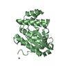
| ||||||||
| Unit cell |
|
-Components
| #1: Protein | Mass: 25853.193 Da / Num. of mol.: 2 Source method: isolated from a genetically manipulated source Source: (gene. exp.) Francisella tularensis subsp. tularensis SCHU S4 (bacteria)Gene: FTT_0507 / Plasmid: pMCSG7 / Production host: #2: Chemical | ChemComp-ZN /   Mass: 65.409 Da / Num. of mol.: 6 / Source method: obtained synthetically / Formula: Zn Mass: 65.409 Da / Num. of mol.: 6 / Source method: obtained synthetically / Formula: Zn#3: Chemical | ChemComp-PEG / |   Mass: 106.120 Da / Num. of mol.: 1 / Source method: obtained synthetically / Formula: C4H10O3 Mass: 106.120 Da / Num. of mol.: 1 / Source method: obtained synthetically / Formula: C4H10O3#4: Water | ChemComp-HOH / |  Mass: 18.015 Da / Num. of mol.: 266 / Source method: isolated from a natural source / Formula: H2O Mass: 18.015 Da / Num. of mol.: 266 / Source method: isolated from a natural source / Formula: H2OHas ligand of interest | N | Has protein modification | N | |
|---|
-Experimental details
-Experiment
| Experiment | Method: X-RAY DIFFRACTION / Number of used crystals: 1 |
|---|
- Sample preparation
| Crystal | Density Matthews: 2.14 Å3/Da / Density % sol: 42.64 % |
|---|---|
| Crystal grow | Temperature: 293 K / Method: vapor diffusion, hanging drop Details: 0.1 M HEPES (pH 7.58), 0.01 mM ZnCl2 and 20% PEG 3350 (v/v), and protein at 58 mg/mL |
-Data collection
| Diffraction | Mean temperature: 100 K / Serial crystal experiment: N |
|---|---|
| Diffraction source | Source: SYNCHROTRON / Site: Australian Synchrotron / Beamline: MX2 / Wavelength: 0.95373 Å |
| Detector | Type: DECTRIS EIGER X 16M / Detector: PIXEL / Date: Apr 14, 2022 |
| Radiation | Protocol: SINGLE WAVELENGTH / Monochromatic (M) / Laue (L): M / Scattering type: x-ray |
| Radiation wavelength | Wavelength: 0.95373 Å / Relative weight: 1 |
| Reflection | Resolution: 1.95→47.39 Å / Num. obs: 31035 / % possible obs: 99.2 % / Redundancy: 4.4 % / CC1/2: 0.996 / Rmerge(I) obs: 0.108 / Rpim(I) all: 0.058 / Rrim(I) all: 0.123 / Net I/σ(I): 8.2 |
| Reflection shell | Resolution: 1.95→2.06 Å / Redundancy: 4.4 % / Rmerge(I) obs: 0.685 / Num. unique obs: 3729 / CC1/2: 0.802 / Rpim(I) all: 0.364 / Rrim(I) all: 0.778 / % possible all: 94.3 |
- Processing
| Software |
| ||||||||||||||||||||||||||||||||||||||||||||||||||||||||||||||||||||||||||||||||||||
|---|---|---|---|---|---|---|---|---|---|---|---|---|---|---|---|---|---|---|---|---|---|---|---|---|---|---|---|---|---|---|---|---|---|---|---|---|---|---|---|---|---|---|---|---|---|---|---|---|---|---|---|---|---|---|---|---|---|---|---|---|---|---|---|---|---|---|---|---|---|---|---|---|---|---|---|---|---|---|---|---|---|---|---|---|---|
| Refinement | Method to determine structure: MOLECULAR REPLACEMENT / Resolution: 1.96→42.2 Å / SU ML: 0.22 / Cross valid method: FREE R-VALUE / σ(F): 1.33 / Phase error: 23.62 / Stereochemistry target values: ML
| ||||||||||||||||||||||||||||||||||||||||||||||||||||||||||||||||||||||||||||||||||||
| Solvent computation | Shrinkage radii: 0.9 Å / VDW probe radii: 1.1 Å / Solvent model: FLAT BULK SOLVENT MODEL | ||||||||||||||||||||||||||||||||||||||||||||||||||||||||||||||||||||||||||||||||||||
| Refinement step | Cycle: LAST / Resolution: 1.96→42.2 Å
| ||||||||||||||||||||||||||||||||||||||||||||||||||||||||||||||||||||||||||||||||||||
| Refine LS restraints |
| ||||||||||||||||||||||||||||||||||||||||||||||||||||||||||||||||||||||||||||||||||||
| LS refinement shell |
| ||||||||||||||||||||||||||||||||||||||||||||||||||||||||||||||||||||||||||||||||||||
| Refinement TLS params. | Method: refined / Origin x: 10.319 Å / Origin y: -8.1251 Å / Origin z: 22.4748 Å
| ||||||||||||||||||||||||||||||||||||||||||||||||||||||||||||||||||||||||||||||||||||
| Refinement TLS group | Selection details: all |
