Movie
Movie Controller


+ Open data

- Basic information
| Entry | Database: PDB / ID: 9edc | ||||||
|---|---|---|---|---|---|---|---|
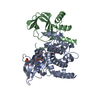
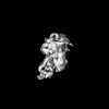
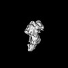
| Title | Reset Type-I Protein Kinase A Holoenzyme | ||||||
 Components Components |
| ||||||
 Keywords Keywords | SIGNALING PROTEIN / Kinase / Regulator | ||||||
| Function / homology |  Function and homology information Function and homology informationPKA-mediated phosphorylation of CREB / PKA-mediated phosphorylation of key metabolic factors / ROBO receptors bind AKAP5 / PKA activation in glucagon signalling / DARPP-32 events / CREB1 phosphorylation through the activation of Adenylate Cyclase / GPER1 signaling / HDL assembly / Factors involved in megakaryocyte development and platelet production / channel activator activity ...PKA-mediated phosphorylation of CREB / PKA-mediated phosphorylation of key metabolic factors / ROBO receptors bind AKAP5 / PKA activation in glucagon signalling / DARPP-32 events / CREB1 phosphorylation through the activation of Adenylate Cyclase / GPER1 signaling / HDL assembly / Factors involved in megakaryocyte development and platelet production / channel activator activity / PKA activation / Regulation of glycolysis by fructose 2,6-bisphosphate metabolism / mitochondrial protein catabolic process / Hedgehog 'off' state / cell communication by electrical coupling involved in cardiac conduction / high-density lipoprotein particle assembly / nucleotide-activated protein kinase complex / Rap1 signalling / potassium channel inhibitor activity / cAMP-dependent protein kinase inhibitor activity / histone H1-4S35 kinase activity / cAMP-dependent protein kinase / regulation of protein processing / cAMP-dependent protein kinase activity / cellular response to parathyroid hormone stimulus / protein localization to lipid droplet / negative regulation of interleukin-2 production / sarcomere organization / regulation of bicellular tight junction assembly / cAMP-dependent protein kinase complex / dorsal/ventral neural tube patterning / Loss of phosphorylation of MECP2 at T308 / CREB1 phosphorylation through the activation of Adenylate Cyclase / PKA activation / Triglyceride catabolism / regulation of osteoblast differentiation / negative regulation of activated T cell proliferation / sperm capacitation / cellular response to cold / High laminar flow shear stress activates signaling by PIEZO1 and PECAM1:CDH5:KDR in endothelial cells / Vasopressin regulates renal water homeostasis via Aquaporins / protein kinase A regulatory subunit binding / negative regulation of glycolytic process through fructose-6-phosphate / ciliary base / protein kinase A catalytic subunit binding / intracellular potassium ion homeostasis / mesoderm formation / RET signaling / Interleukin-3, Interleukin-5 and GM-CSF signaling / immunological synapse / PKA activation in glucagon signalling / sperm head-tail coupling apparatus / regulation of cardiac conduction / plasma membrane raft / Regulation of MECP2 expression and activity / DARPP-32 events / regulation of macroautophagy / cAMP/PKA signal transduction / cardiac muscle cell proliferation / axoneme / regulation of cardiac muscle contraction / sperm flagellum / postsynaptic modulation of chemical synaptic transmission / cAMP binding / vascular endothelial cell response to laminar fluid shear stress / renal water homeostasis / negative regulation of cAMP/PKA signal transduction / Hedgehog 'off' state / multivesicular body / Ion homeostasis / negative regulation of protein localization to chromatin / negative regulation of TORC1 signaling / regulation of cardiac muscle contraction by regulation of the release of sequestered calcium ion / protein serine/threonine/tyrosine kinase activity / Loss of Nlp from mitotic centrosomes / Loss of proteins required for interphase microtubule organization from the centrosome / regulation of proteasomal protein catabolic process / Recruitment of mitotic centrosome proteins and complexes / cellular response to epinephrine stimulus / protein export from nucleus / cellular response to glucagon stimulus / Recruitment of NuMA to mitotic centrosomes / calcium channel complex / Anchoring of the basal body to the plasma membrane / CD209 (DC-SIGN) signaling / Mitochondrial protein degradation / regulation of heart rate / positive regulation of phagocytosis / FCGR3A-mediated IL10 synthesis / positive regulation of calcium-mediated signaling / positive regulation of gluconeogenesis / acrosomal vesicle / AURKA Activation by TPX2 / positive regulation of protein export from nucleus / regulation of microtubule cytoskeleton organization / negative regulation of smoothened signaling pathway / neuromuscular junction / neural tube closure / Regulation of insulin secretion / cellular response to glucose stimulus Similarity search - Function | ||||||
| Biological species |  Homo sapiens (human) Homo sapiens (human) | ||||||
| Method | ELECTRON MICROSCOPY / single particle reconstruction / cryo EM / Resolution: 4.18 Å | ||||||
 Authors Authors | Venkatakrishnan, V. / Buckley, T. / Laremore, T.N. / Armache, J.P. / Anand, G.S. | ||||||
| Funding support |  United States, 1items United States, 1items
| ||||||
 Citation Citation | Journal: J Am Chem Soc / Year: 2025 Title: Multiplicity of Regulatory Subunit Conformations Defines Structural Ensemble of Reset Protein Kinase A Holoenzyme. Authors: Varun Venkatakrishnan / Tatiana N Laremore / Theresa S C Buckley / Jean-Paul Armache / Ganesh S Anand / Abstract: How protein kinase A (PKA) is reset to a basal state following 3'5'-cyclic adenosine monophosphate (cAMP)-mediated activation is unknown. Here we describe the mechanism of cAMP-PKA type I signal ...How protein kinase A (PKA) is reset to a basal state following 3'5'-cyclic adenosine monophosphate (cAMP)-mediated activation is unknown. Here we describe the mechanism of cAMP-PKA type I signal termination leading to a reset of PKA by holoenzyme formation through the obligatory action of phosphodiesterases (PDEs). We report a catalytic subunit (Cα)-assisted mechanism for the reset of type I PKA and describe for the first time multiple structures of the reset PKA holoenzyme (RIα:Cα) that capture an ensemble of multiple conformational end-states through integrative electron microscopy and structural mass spectrometry approaches. Together these complementary methods highlight the large conformational dynamics of the regulatory subunit (RIα) within the tetrameric reset PKA holoenzyme. The cAMP-free reset PKA holoenzyme adopts multiple distinct conformations of RIα with contributions from the N-terminal linker and CNB-B dynamics. Our findings highlight the interplay between RIα, Cα, and PDEs (PDE8) in cAMP-PKA signalosomes to offer a new paradigm for PDE-mediated regulation of cAMP-PKA signaling. | ||||||
| History |
|
- Structure visualization
| Structure viewer | Molecule: MolmilJmol/JSmol |
|---|
- Downloads & links
-Download
| PDBx/mmCIF format | 9edc.cif.gz | 111.2 KB | Display | PDBx/mmCIF format |
|---|---|---|---|---|
| PDB format | pdb9edc.ent.gz | 79.6 KB | Display | PDB format |
| PDBx/mmJSON format | 9edc.json.gz | Tree view | PDBx/mmJSON format | |
| Others |  Other downloads Other downloads |
-Validation report
| Arichive directory | https://data.pdbj.org/pub/pdb/validation_reports/ed/9edcftp://data.pdbj.org/pub/pdb/validation_reports/ed/9edc | HTTPS FTP |
|---|
-Related structure data
| Related structure data | 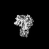 47944MC 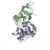 9eddC 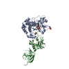 9edeC M: map data used to model this data C: citing same article ( |
|---|---|
| Similar structure data |
-Links
 PDBj
PDBj


- Assembly
| Deposited unit | 
|
|---|---|
| 1 |
|
-Components
| #1: Protein | Mass: 40737.297 Da / Num. of mol.: 1 Source method: isolated from a genetically manipulated source Source: (gene. exp.) Homo sapiens (human) / Gene: PRKACA, PKACA / Production host:  |
|---|---|
| #2: Protein | Mass: 47415.410 Da / Num. of mol.: 1 Source method: isolated from a genetically manipulated source Source: (gene. exp.) |
| #3: Chemical | ChemComp-ATP /   Mass: 507.181 Da / Num. of mol.: 1 / Source method: obtained synthetically / Formula: C10H16N5O13P3 / Feature type: SUBJECT OF INVESTIGATION / Comment: ATP, energy-carrying molecule*YM Mass: 507.181 Da / Num. of mol.: 1 / Source method: obtained synthetically / Formula: C10H16N5O13P3 / Feature type: SUBJECT OF INVESTIGATION / Comment: ATP, energy-carrying molecule*YM |
| Has ligand of interest | Y |
| Has protein modification | Y |
-Experimental details
-Experiment
| Experiment | Method: ELECTRON MICROSCOPY |
|---|---|
| EM experiment | Aggregation state: PARTICLE / 3D reconstruction method: single particle reconstruction |
- Sample preparation
| Component | Name: Reset type-I PKA holoenzyme complex of regulatory and catalytic subunits Type: COMPLEX / Entity ID: #1-#2 / Source: RECOMBINANT | ||||||||||||||||||||||||||||||
|---|---|---|---|---|---|---|---|---|---|---|---|---|---|---|---|---|---|---|---|---|---|---|---|---|---|---|---|---|---|---|---|
| Molecular weight | Experimental value: NO | ||||||||||||||||||||||||||||||
| Source (natural) | Organism: Homo sapiens (human) | ||||||||||||||||||||||||||||||
| Source (recombinant) | Organism: | ||||||||||||||||||||||||||||||
| Buffer solution | pH: 7 | ||||||||||||||||||||||||||||||
| Buffer component |
| ||||||||||||||||||||||||||||||
| Specimen | Conc.: 0.6 mg/ml / Embedding applied: NO / Shadowing applied: NO / Staining applied: NO / Vitrification applied: YES | ||||||||||||||||||||||||||||||
| Specimen support | Grid type: Quantifoil R2/1 | ||||||||||||||||||||||||||||||
| Vitrification | Instrument: FEI VITROBOT MARK IV / Cryogen name: ETHANE |
- Electron microscopy imaging
| Experimental equipment |  Model: Talos Arctica / Image courtesy: FEI Company |
|---|---|
| Microscopy | Model: FEI TALOS ARCTICA |
| Electron gun | Electron source:  FIELD EMISSION GUN / Accelerating voltage: 200 kV / Illumination mode: SPOT SCAN FIELD EMISSION GUN / Accelerating voltage: 200 kV / Illumination mode: SPOT SCAN |
| Electron lens | Mode: OTHER / Nominal defocus max: 2200 nm / Nominal defocus min: 1000 nm |
| Image recording | Electron dose: 49.66 e/Å2 / Film or detector model: FEI FALCON IV (4k x 4k) / Num. of real images: 4559 |
- Processing
| EM software |
| ||||||||||||||||||||||||||||||||||||||||
|---|---|---|---|---|---|---|---|---|---|---|---|---|---|---|---|---|---|---|---|---|---|---|---|---|---|---|---|---|---|---|---|---|---|---|---|---|---|---|---|---|---|
| CTF correction | Type: PHASE FLIPPING AND AMPLITUDE CORRECTION | ||||||||||||||||||||||||||||||||||||||||
| Particle selection | Num. of particles selected: 2683140 | ||||||||||||||||||||||||||||||||||||||||
| Symmetry | Point symmetry: C1 (asymmetric) | ||||||||||||||||||||||||||||||||||||||||
| 3D reconstruction | Resolution: 4.18 Å / Resolution method: FSC 0.143 CUT-OFF / Num. of particles: 95294 / Symmetry type: POINT | ||||||||||||||||||||||||||||||||||||||||
| Atomic model building | B value: 369.72 / Protocol: RIGID BODY FIT / Space: REAL | ||||||||||||||||||||||||||||||||||||||||
| Atomic model building |
| ||||||||||||||||||||||||||||||||||||||||
| Refine LS restraints |
|