Movie
Movie Controller


+ Open data

- Basic information
| Entry | Database: PDB / ID: 9.0E+96 | |||||||||||||||||||||||||||||||||||||||
|---|---|---|---|---|---|---|---|---|---|---|---|---|---|---|---|---|---|---|---|---|---|---|---|---|---|---|---|---|---|---|---|---|---|---|---|---|---|---|---|---|
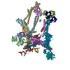
| Title | WEEV CBA87 VLP in complex with human PCDH10-EC1 | |||||||||||||||||||||||||||||||||||||||
 Components Components |
| |||||||||||||||||||||||||||||||||||||||
 Keywords Keywords | VIRAL PROTEIN / Alphavirus Receptor | |||||||||||||||||||||||||||||||||||||||
| Function / homology |  Function and homology information Function and homology informationtogavirin / T=4 icosahedral viral capsid / host cell endoplasmic reticulum / homophilic cell-cell adhesion / cell adhesion molecule binding / nervous system development / channel activity / monoatomic ion transmembrane transport / symbiont-mediated suppression of host toll-like receptor signaling pathway / host cell cytoplasm ...togavirin / T=4 icosahedral viral capsid / host cell endoplasmic reticulum / homophilic cell-cell adhesion / cell adhesion molecule binding / nervous system development / channel activity / monoatomic ion transmembrane transport / symbiont-mediated suppression of host toll-like receptor signaling pathway / host cell cytoplasm / host cell Golgi apparatus / entry receptor-mediated virion attachment to host cell / postsynaptic membrane / cell adhesion / symbiont-mediated suppression of host gene expression / serine-type endopeptidase activity / fusion of virus membrane with host endosome membrane / calcium ion binding / symbiont entry into host cell / virion attachment to host cell / host cell nucleus / host cell plasma membrane / virion membrane / structural molecule activity / glutamatergic synapse / proteolysis / RNA binding / plasma membrane Similarity search - Function | |||||||||||||||||||||||||||||||||||||||
| Biological species |  Western equine encephalitis virus Western equine encephalitis virus Homo sapiens (human) Homo sapiens (human) | |||||||||||||||||||||||||||||||||||||||
| Method | ELECTRON MICROSCOPY / single particle reconstruction / cryo EM / Resolution: 4.05 Å | |||||||||||||||||||||||||||||||||||||||
 Authors Authors | Raju, S. / Diamond, M.S. / Fremont, D.H. / Center for Structural Biology of Infectious Diseases (CSBID) | |||||||||||||||||||||||||||||||||||||||
| Funding support |  United States, 1items United States, 1items
| |||||||||||||||||||||||||||||||||||||||
 Citation Citation | Journal: Cell / Year: 2025 Title: Structural basis for plasticity in receptor engagement by an encephalitic alphavirus. Authors: Saravanan Raju / Sathvik Palakurty / Alan Sariol / Ngan Wagoner / Lucas J Adams / Sean Hui / William B Klimstra / Daved H Fremont / Michael S Diamond / Abstract: The structural basis for shifts in receptor usage remains poorly understood despite the implications for virus adaptation and emergence. Western equine encephalitis virus (WEEV) strains exhibit ...The structural basis for shifts in receptor usage remains poorly understood despite the implications for virus adaptation and emergence. Western equine encephalitis virus (WEEV) strains exhibit different patterns of engagement for two of their entry receptors: very-low-density lipoprotein receptor (VLDLR) and protocadherin 10 (PCDH10). Using structural and functional studies, we show that while all WEEV strains have a lipoprotein class A (LA) domain binding site near the E1 fusion loop, VLDLR engagement requires a second binding site in E2 that can vary with single nucleotide substitutions. We also resolve a structure of PCDH10 bound to WEEV, which reveals interactions near the E1 fusion loop with residues that also mediate LA domain binding. Evolutionary analysis enabled the generation of a PCDH10 decoy that protects in vivo against all WEEV strains tested. Our experiments demonstrate how viruses can engage multiple receptors using shared determinants, which likely impacts cellular tropism and virulence. | |||||||||||||||||||||||||||||||||||||||
| History |
|
- Structure visualization
| Structure viewer | Molecule: MolmilJmol/JSmol |
|---|
- Downloads & links
-Download
| PDBx/mmCIF format | 9e96.cif.gz | 748.1 KB | Display | PDBx/mmCIF format |
|---|---|---|---|---|
| PDB format | pdb9e96.ent.gz | 614.3 KB | Display | PDB format |
| PDBx/mmJSON format | 9e96.json.gz | Tree view | PDBx/mmJSON format | |
| Others |  Other downloads Other downloads |
-Validation report
| Arichive directory | https://data.pdbj.org/pub/pdb/validation_reports/e9/9e96ftp://data.pdbj.org/pub/pdb/validation_reports/e9/9e96 | HTTPS FTP |
|---|
-Related structure data
| Related structure data |  47775MC  9e9yC  9e9zC  9eauC M: map data used to model this data C: citing same article ( |
|---|---|
| Similar structure data |


-Links
 PDBj
PDBj


- Assembly
| Deposited unit | 
|
|---|---|
| 1 |
|
-Components
| #1: Protein | Mass: 47345.758 Da / Num. of mol.: 4 Source method: isolated from a genetically manipulated source Source: (gene. exp.) Western equine encephalitis virus / Production host: Homo sapiens (human) / References: UniProt: Q1W679#2: Protein | Mass: 45393.867 Da / Num. of mol.: 4 Source method: isolated from a genetically manipulated source Source: (gene. exp.) Western equine encephalitis virus / Strain: CBA87 / Production host: Homo sapiens (human) / References: UniProt: Q1W679#3: Protein | Mass: 16712.816 Da / Num. of mol.: 4 Source method: isolated from a genetically manipulated source Source: (gene. exp.) Western equine encephalitis virus / Production host: Homo sapiens (human) / References: UniProt: P13897, togavirin#4: Protein | Mass: 11866.213 Da / Num. of mol.: 4 Source method: isolated from a genetically manipulated source Source: (gene. exp.) Homo sapiens (human) / Gene: PCDH10, KIAA1400 / Production host: Homo sapiens (human) / References: UniProt: Q9P2E7Has protein modification | Y | |
|---|
-Experimental details
-Experiment
| Experiment | Method: ELECTRON MICROSCOPY |
|---|---|
| EM experiment | Aggregation state: PARTICLE / 3D reconstruction method: single particle reconstruction |
- Sample preparation
| Component | Name: Western equine encephalitis virus / Type: VIRUS Details: virus-like particles were produced recombinantly in 293T cells and purified by PEG precipitation followed by density gradient ultracentrifugation. Entity ID: all / Source: MULTIPLE SOURCES |
|---|---|
| Source (natural) | Organism: Western equine encephalitis virus / Strain: CBA87 |
| Source (recombinant) | Organism: Homo sapiens (human) |
| Details of virus | Empty: YES / Enveloped: YES / Isolate: STRAIN / Type: VIRUS-LIKE PARTICLE |
| Natural host | Organism: Passeridae |
| Buffer solution | pH: 7.4 |
| Specimen | Embedding applied: NO / Shadowing applied: NO / Staining applied: NO / Vitrification applied: YES |
| Vitrification | Cryogen name: ETHANE |
- Electron microscopy imaging
| Experimental equipment |  Model: Titan Krios / Image courtesy: FEI Company |
|---|---|
| Microscopy | Model: TFS KRIOS |
| Electron gun | Electron source:  FIELD EMISSION GUN / Accelerating voltage: 300 kV / Illumination mode: FLOOD BEAM FIELD EMISSION GUN / Accelerating voltage: 300 kV / Illumination mode: FLOOD BEAM |
| Electron lens | Mode: BRIGHT FIELD / Nominal defocus max: 2100 nm / Nominal defocus min: 700 nm |
| Image recording | Electron dose: 52 e/Å2 / Film or detector model: FEI FALCON IV (4k x 4k) |
- Processing
| EM software |
| ||||||||||||||||
|---|---|---|---|---|---|---|---|---|---|---|---|---|---|---|---|---|---|
| CTF correction | Type: PHASE FLIPPING AND AMPLITUDE CORRECTION | ||||||||||||||||
| 3D reconstruction | Resolution: 4.05 Å / Resolution method: FSC 0.143 CUT-OFF / Num. of particles: 231651 / Symmetry type: POINT | ||||||||||||||||
| Atomic model building | PDB-ID: 8DEE Accession code: 8DEE / Source name: PDB / Type: experimental model | ||||||||||||||||
| Refinement | Highest resolution: 4.05 Å Stereochemistry target values: REAL-SPACE (WEIGHTED MAP SUM AT ATOM CENTERS) |