 Movie
Movie Controller

[English] 日本語
 Yorodumi
- PDB-9e37: Polaromonas naphthalenivorans phosphoenolpyruvate carboxykinase i... -
+ Open data

- Basic information
| Entry | Database: PDB / ID: 9.0E+37 | ||||||
|---|---|---|---|---|---|---|---|
| Title | Polaromonas naphthalenivorans phosphoenolpyruvate carboxykinase in complex with oxalate and GTP | ||||||
 Components Components | Phosphoenolpyruvate carboxykinase [GTP] | ||||||
 Keywords Keywords | LYASE / Inhibitor complex / Metabolic enzyme / Multi-temperature / Ambient temperature | ||||||
| Function / homology |  Function and homology information Function and homology informationphosphoenolpyruvate carboxykinase (GTP) / phosphoenolpyruvate carboxykinase (GTP) activity / : / propionate catabolic process / oxaloacetate metabolic process / response to starvation / response to lipid / gluconeogenesis / cellular response to glucose stimulus / kinase activity ...phosphoenolpyruvate carboxykinase (GTP) / phosphoenolpyruvate carboxykinase (GTP) activity / : / propionate catabolic process / oxaloacetate metabolic process / response to starvation / response to lipid / gluconeogenesis / cellular response to glucose stimulus / kinase activity / manganese ion binding / GTP binding / cytosol Similarity search - Function | ||||||
| Biological species |  Polaromonas naphthalenivorans (bacteria) Polaromonas naphthalenivorans (bacteria) | ||||||
| Method |  X-RAY DIFFRACTION / MOLECULAR REPLACEMENT / Resolution: 2 Å X-RAY DIFFRACTION / MOLECULAR REPLACEMENT / Resolution: 2 Å | ||||||
 Authors Authors | McLeod, M.J. / Holyoak, T. | ||||||
| Funding support |  Canada, 1items Canada, 1items
| ||||||
 Citation Citation | Journal: Protein Sci. / Year: 2025 Title: Structural mechanisms for cold-adapted activity of phosphoenolpyruvate carboxykinase. Authors: McLeod, M.J. / Yazdani, S. / Barwell, S.A.E. / Holyoak, T. | ||||||
| History |
|
- Structure visualization
| Structure viewer | Molecule: MolmilJmol/JSmol |
|---|
- Downloads & links
-Download
| PDBx/mmCIF format | 9e37.cif.gz | 182.1 KB | Display | PDBx/mmCIF format |
|---|---|---|---|---|
| PDB format | pdb9e37.ent.gz | 112 KB | Display | PDB format |
| PDBx/mmJSON format | 9e37.json.gz | Tree view | PDBx/mmJSON format | |
| Others |  Other downloads Other downloads |
-Validation report
| Arichive directory | https://data.pdbj.org/pub/pdb/validation_reports/e3/9e37ftp://data.pdbj.org/pub/pdb/validation_reports/e3/9e37 | HTTPS FTP |
|---|
-Related structure data
| Related structure data |  9e32C  9e33C  9e34C  9e35C  9e36C  9e38C C: citing same article ( |
|---|---|
| Similar structure data |
-Links
 PDBj
PDBj- Assembly
| Deposited unit | 
| ||||||||||||
|---|---|---|---|---|---|---|---|---|---|---|---|---|---|
| 1 |
| ||||||||||||
| Unit cell |
|
-Components
-Protein , 1 types, 1 molecules A
| #1: Protein | Mass: 68353.320 Da / Num. of mol.: 1 Source method: isolated from a genetically manipulated source Source: (gene. exp.) Polaromonas naphthalenivorans (bacteria)Gene: pckG, Pnap_0102, Pck1 / Production host: References: UniProt: A1VIE9, phosphoenolpyruvate carboxykinase (GTP) |
|---|
-Non-polymers , 7 types, 515 molecules 


| #2: Chemical | ChemComp-GTP /  Mass: 523.180 Da / Num. of mol.: 1 / Source method: obtained synthetically / Formula: C10H16N5O14P3 / Feature type: SUBJECT OF INVESTIGATION / Comment: GTP, energy-carrying molecule*YM Mass: 523.180 Da / Num. of mol.: 1 / Source method: obtained synthetically / Formula: C10H16N5O14P3 / Feature type: SUBJECT OF INVESTIGATION / Comment: GTP, energy-carrying molecule*YM |
|---|---|
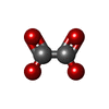
| #3: Chemical | ChemComp-OXL /  Mass: 88.019 Da / Num. of mol.: 1 / Source method: obtained synthetically / Formula: C2O4 / Feature type: SUBJECT OF INVESTIGATION Mass: 88.019 Da / Num. of mol.: 1 / Source method: obtained synthetically / Formula: C2O4 / Feature type: SUBJECT OF INVESTIGATION |
| #4: Chemical | ChemComp-MN /  Mass: 54.938 Da / Num. of mol.: 1 / Source method: obtained synthetically / Formula: Mn Mass: 54.938 Da / Num. of mol.: 1 / Source method: obtained synthetically / Formula: Mn |
| #5: Chemical | ChemComp-MG /  Mass: 24.305 Da / Num. of mol.: 1 / Source method: obtained synthetically / Formula: Mg Mass: 24.305 Da / Num. of mol.: 1 / Source method: obtained synthetically / Formula: Mg |
| #6: Chemical | ChemComp-PGE /  Mass: 150.173 Da / Num. of mol.: 1 / Source method: obtained synthetically / Formula: C6H14O4 Mass: 150.173 Da / Num. of mol.: 1 / Source method: obtained synthetically / Formula: C6H14O4 |
| #7: Chemical | ChemComp-1PE /  Mass: 238.278 Da / Num. of mol.: 1 / Source method: obtained synthetically / Formula: C10H22O6 / Comment: precipitant*YM Mass: 238.278 Da / Num. of mol.: 1 / Source method: obtained synthetically / Formula: C10H22O6 / Comment: precipitant*YM |
| #8: Water | ChemComp-HOH / Mass: 18.015 Da / Num. of mol.: 509 / Source method: isolated from a natural source / Formula: H2O |
-Details
| Has ligand of interest | Y |
|---|---|
| Has protein modification | N |
-Experimental details
-Experiment
| Experiment | Method: X-RAY DIFFRACTION / Number of used crystals: 1 |
|---|
- Sample preparation
| Crystal | Density Matthews: 2.42 Å3/Da / Density % sol: 49.15 % |
|---|---|
| Crystal grow | Temperature: 293 K / Method: vapor diffusion, hanging drop / pH: 8 Details: 100 mM TRIS-Cl pH 8.0, 0.4 M LiCl2, 5 mM MgCl2, 1 mM MnCl2, 30-40% PEG 8000, 10 mM oxalate and 10 mM GTP. 15 mg/mL PnPEPCK |
-Data collection
| Diffraction | Mean temperature: 100 K / Serial crystal experiment: N |
|---|---|
| Diffraction source | Source: ROTATING ANODE / Type: RIGAKU / Wavelength: 1.54 Å |
| Detector | Type: RIGAKU / Detector: IMAGE PLATE / Date: Jan 1, 2023 |
| Radiation | Protocol: SINGLE WAVELENGTH / Monochromatic (M) / Laue (L): M / Scattering type: x-ray |
| Radiation wavelength | Wavelength: 1.54 Å / Relative weight: 1 |
| Reflection | Resolution: 2→30.5 Å / Num. obs: 49489 / % possible obs: 98.9 % / Redundancy: 13.1 % / Biso Wilson estimate: 30.6 Å2 / CC1/2: 0.962 / CC star: 0.999 / Rmerge(I) obs: 0.119 / Rpim(I) all: 0.033 / Rrim(I) all: 0.124 / Net I/σ(I): 13.8 |
| Reflection shell | Resolution: 2→2.07 Å / Redundancy: 6.7 % / Rmerge(I) obs: 0.637 / Mean I/σ(I) obs: 2.8 / Num. unique obs: 4114 / CC1/2: 0.824 / CC star: 0.95 / Rpim(I) all: 0.244 / Rrim(I) all: 0.686 / % possible all: 91.2 |
- Processing
| Software |
| ||||||||||||||||||||||||||||||||||||||||||||||||||||||||||||||||||||||||||||||||||||||||||||||||||||||||||||||||
|---|---|---|---|---|---|---|---|---|---|---|---|---|---|---|---|---|---|---|---|---|---|---|---|---|---|---|---|---|---|---|---|---|---|---|---|---|---|---|---|---|---|---|---|---|---|---|---|---|---|---|---|---|---|---|---|---|---|---|---|---|---|---|---|---|---|---|---|---|---|---|---|---|---|---|---|---|---|---|---|---|---|---|---|---|---|---|---|---|---|---|---|---|---|---|---|---|---|---|---|---|---|---|---|---|---|---|---|---|---|---|---|---|---|
| Refinement | Method to determine structure: MOLECULAR REPLACEMENT / Resolution: 2→30.5 Å / SU ML: 0.2362 / Cross valid method: FREE R-VALUE / σ(F): 1.34 / Phase error: 20.4407 Stereochemistry target values: GeoStd + Monomer Library + CDL v1.2
| ||||||||||||||||||||||||||||||||||||||||||||||||||||||||||||||||||||||||||||||||||||||||||||||||||||||||||||||||
| Solvent computation | Shrinkage radii: 0.9 Å / VDW probe radii: 1.1 Å / Solvent model: FLAT BULK SOLVENT MODEL | ||||||||||||||||||||||||||||||||||||||||||||||||||||||||||||||||||||||||||||||||||||||||||||||||||||||||||||||||
| Displacement parameters | Biso mean: 32.59 Å2 | ||||||||||||||||||||||||||||||||||||||||||||||||||||||||||||||||||||||||||||||||||||||||||||||||||||||||||||||||
| Refinement step | Cycle: LAST / Resolution: 2→30.5 Å
| ||||||||||||||||||||||||||||||||||||||||||||||||||||||||||||||||||||||||||||||||||||||||||||||||||||||||||||||||
| Refine LS restraints |
| ||||||||||||||||||||||||||||||||||||||||||||||||||||||||||||||||||||||||||||||||||||||||||||||||||||||||||||||||
| LS refinement shell |
|
